Interactive figure legend
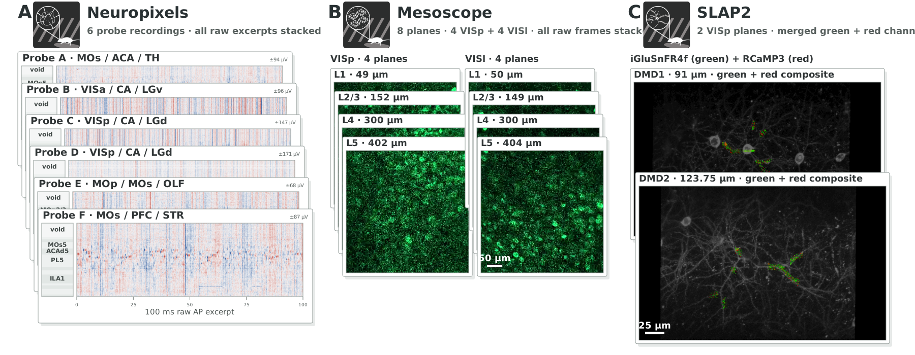
Representative extracellular recordings from six Neuropixels probes. The voltage map shows recording depth and sampled anatomical regions; colored markers identify spikes assigned to sorted units. The selected unit's activity is reported as firing rate (spikes/s), alongside its mean spike template. Common-mode correction removes the across-contact median at each time point.
Representative calcium recordings from eight mesoscope imaging planes in VISp and VISl. Raw images are unprocessed fluorescence frames; registered movies are motion corrected and decrosstalked. The reference is the registered mean image. Colored outlines identify extracted regions of interest, with the selected region's activity reported as ΔF/F (%).
Representative SLAP2 recordings from two imaging depths. Raw movies show individual iGluSnFR4f or RCaMP3 detector samples from single acquisition cycles, with unsampled pixels black and no reference-image background. Registered movies and extracted source traces use iGluSnFR4f; the reference is the registered mean image. Outlines identify extracted sources; source counts are not neuron counts. Source activity is reported as ΔF/F (%), including when the raw RCaMP3 channel is displayed.
Segmentation masks use registered coordinates and are not overlaid on raw frames. Movie and trace excerpts have separate time axes.