VIP function is not uniform across the brain. The disinhibitory VIP→SST→Pyr disinhibitory motif first formalised by Pi et al., 2013 in auditory cortex and medial prefrontal cortex captures a real and reproducible circuit, but contemporary multi-area recordings show that the operation implemented by that motif — and in some structures, the motif itself — varies systematically with cortical area, behavioural context, and developmental origin Pi et al., 2013Karnani et al., 2016Dipoppa et al., 2018Lee et al., 2013Bastos et al., 2023. Primary visual cortex emphasises locomotion-gated, contrast-dependent gain enhancement Dipoppa et al., 2018Millman et al., 2020Pakan et al., 2016; auditory cortex deploys VIP cells for reinforcement-linked disinhibition and critical-period plasticity Pi et al., 2013; somatosensory cortex recruits them via motor copy during active sensing Lee et al., 2013Sermet et al., 2019; medial prefrontal and anterior cingulate cortex implement top-down control of attention, working memory, and pain Bastos et al., 2023Anastasiades et al., 2021Kamigaki & Dan, 2017Li et al., 2022; and hippocampal VIP/calretinin “IS-3” cells assemble a circuit motif distinct from neocortex, targeting other interneurons rather than principal cells Gulyás et al., 1996Tyan et al., 2014Tricoire et al., 2011. Subcortical VIP populations — in basolateral amygdala, dorsal striatum, and the suprachiasmatic nucleus — extend the diversity further, including a non-GABAergic, peptidergic projection role in circadian timekeeping Rhomberg et al., 2018Vereczki et al., 2021Muñoz-Manchado et al., 2018Maywood et al., 2006Cutler et al., 2003. These cross-region differences place strong constraints on any unitary theory of VIP function and motivate the area-by-area treatment that follows. We refer to the previous section’s behavioural heterogeneity (In Vivo Function During Behavior) and forward to the temporal-coordination signatures examined in Oscillatory Dynamics and Temporal Coordination.
Primary visual cortex: locomotion gating and contrast-dependent gain
The most extensively characterised cortical VIP population sits in mouse V1, where VIP+ small bipolar/multipolar cells with vertically descending axonal arbors target apical dendrites of L5 pyramidal neurons and innervate L2/3 SST cells Kawaguchi & Kubota, 1996Gonchar, 2008Kawaguchi, 1997. Long-range tracing places these cells at the receiving end of cholinergic, serotonergic, and frontal cortical inputs disproportionately enriched relative to PV/SST cells Wall et al., 2016. Two-photon imaging during head-fixed running has converged on a basic phenomenology: VIP cells in V1 show the strongest run-related rise of any genetically defined interneuron class, while SST cells move in the opposite direction, and PV cells track stimulus contrast more than locomotion Dipoppa et al., 2018Pakan et al., 2016Karnani et al., 2016. The resulting state-dependent VIP→SST motif underlies a multiplicative or additive rescaling of pyramidal responses that is widely interpreted as locomotion-driven gain enhancement Pi et al., 2013Dipoppa et al., 2018Karnani et al., 2016Millman et al., 2020.
Three lines of evidence complicate this disinhibitory reading. First, the locomotion drive itself is context-modulated: Pakan et al. (2016) reported that VIP↑/SST↓ antiphasic modulation in V1 is most pronounced when concurrent visual stimuli are present, although most VIP cells remain locomotion-responsive in darkness, in agreement with the more invariant locomotion drive reported by Dipoppa et al. (2018). Second, the gain modulation is not a uniform multiplicative rescaling: Millman et al. (2020) showed that V1 VIP activation enhances responses preferentially at low contrasts and lowers contrast threshold without changing peak firing at saturating contrasts, and that this contrast-dependent enhancement requires intact SST inhibition. Third, the spatial structure of disinhibition is not blanket: Karnani et al. (2016) documented a localised “spotlight” of VIP-mediated disinhibition that opens spatially restricted holes in SST inhibition rather than uniformly suppressing it. These findings together argue that V1 VIP cells implement a contextual, spatially structured, contrast-dependent operation rather than the uniform broadcast disinhibition assumed by the textbook motif.
The morphological and laminar substrate constrains how broadly V1 VIP cells can act. Gonchar (2008) documented that L2/3 small bipolar VIP cells display dense vertical axonal arbors descending to L5, consistent with apical-dendrite-targeting onto pyramidal neurons in addition to L2/3 SST cells, and Kawaguchi & Kubota (1996) and Kawaguchi (1997) established that the bipolar/double-bouquet morphology distinguishes VIP cells from PV multipolar baskets and SST Martinotti cells Kawaguchi & Kubota, 1996Kawaguchi, 1997Gonchar, 2008. The cells therefore have direct anatomical access to apical dendrites of L5 pyramidal neurons, providing one route by which top-down signals from cingulate cortex can be funnelled into deep-layer pyramidal output via VIP recruitment Bastos et al., 2023Wall et al., 2016. Differential thalamocortical drive is also relevant: Ji et al. (2015) reported broadly similar laminar innervation patterns for auditory and visual thalamocortical pathways onto cortical interneurons, with quantitative variation across interneuron classes — including VIP — that is consistent with the input-specificity differences seen across cortical areas Wall et al., 2016Sermet et al., 2019.
V1 VIP cells are also a substrate for top-down context. Cingulate-to-V1 long-range projections preferentially synapse onto a subset of V1 VIP cells, and optogenetic silencing of those projections degrades figure–ground discrimination behaviourally. The targeting bias is consistent with the rabies-tracing analysis of Wall et al. (2016), which showed that VIP cells across cortex receive disproportionately enriched long-range input from frontal, neuromodulatory, and thalamic sources relative to PV/SST cells Wall et al., 2016Anastasiades et al., 2021. The morphological substrate identified by Kawaguchi & Kubota (1996) and Gonchar (2008) — bipolar VIP cells with vertical apical-targeting axons — provides the direct anatomical route from this top-down input onto deep-layer pyramidal output Kawaguchi & Kubota, 1996Gonchar, 2008Bastos et al., 2023. This anchors the V1 motif to broader cortical hierarchies and links it to the frontal subsections below (In Vivo Function During Behavior).
Auditory cortex: reinforcement, plasticity gating, and the polarity puzzle
Auditory cortex is the disinhibitory home of the VIP→SST→Pyr motif. Pi et al. (2013) performed the foundational experiment: optogenetic activation of A1 VIP cells transiently suppressed primarily SST and a fraction of PV interneurons, disinhibiting pyramidal cells, and silencing VIP cells reduced pyramidal firing during reinforcement, identifying VIP cells as a state-dependent gain mechanism activated by reward and punishment Pi et al., 2013. The motif is layer-biased, with strongest disinhibitory effects in L2/3, and is supported by convergent long-range cortical and subcortical drive consistent with VIP cells being broadcast targets of neuromodulatory and top-down signals Pi et al., 2013Wall et al., 2016.
Two extensions of the disinhibitory A1 motif organise much of the subsequent literature. First, BLA VIP cells implement adaptive disinhibition during associative learning: Krabbe et al. (2019) showed that VIP activity in basolateral amygdala grows progressively across fear-conditioning sessions, that VIP cells acquire CS+/CS− selectivity, and that silencing them during conditioning impairs memory consolidation, identifying disinhibition itself as a plastic substrate rather than a static gain Krabbe et al., 2019. The drive into these cells comes from the basal nucleus and thalamus, providing an arousal-linked substrate for state-dependent recruitment Krabbe et al., 2019Wall et al., 2016. Second, a layer-1 microcircuit gates auditory critical-period plasticity: Takesian et al. (2018) showed that L1 interneurons receive nicotinic cholinergic input from basal forebrain and inhibit L2/3 PV cells, and that recruiting these L1 cells in adults reopens tonotopic plasticity — extending the motif from instantaneous gain to developmental and recovery time-scales Takesian et al., 2018.
Auditory cortex is also where the cross-area “polarity puzzle” becomes acute. In V1, locomotion drives VIP cells up and improves visual encoding Dipoppa et al., 2018Pakan et al., 2016; in A1, the parallel literature reports that locomotion suppresses spiking and reduces encoding efficiency, opposite in sign to V1, despite the apparently identical disinhibitory motif. The keys reporting that A1 polarity reversal directly (Fu 2014; Bigelow 2019) lie in the temporal-coordination cluster covered in Oscillatory Dynamics and Temporal Coordination and are not present in this section’s filtered citation set; we flag this as an unmet citation need (see Caveats and unmet citation needs below). Within the present packet, the available evidence shows that A1 VIP cells are robustly recruited by reinforcement and learning drives Pi et al., 2013Takesian et al., 2018Wall et al., 2016 rather than by locomotion per se, which is consistent with — but does not in isolation prove — an A1/V1 polarity divergence in locomotion gating relative to V1 Dipoppa et al., 2018Pakan et al., 2016.
Somatosensory cortex: motor copy and orthogonal attention
Barrel cortex provides the clearest example of motor-context gating of VIP cells. Lee et al. (2013) showed that whisking-induced motor inputs from M1 activate S1 VIP cells, which inhibit SST cells and disinhibit L2/3 pyramidal neurons during active sensing; optogenetic VIP silencing during active whisking reduced pyramidal firing while VIP activation mimicked the whisking-induced enhancement, establishing the VIP→SST motif as disinhibitory in S1 as well as A1 and mPFC Lee et al., 2013Pi et al., 2013. Importantly, the recruiting input is the motor command, not passive whisker deflection — VIP cells in S1 respond to what the animal is doing rather than only to what it senses Lee et al., 2013.
Thalamic input specificity refines this picture. Sermet et al. (2019) mapped POm and VPM inputs and found that S1 VIP cells receive intermediate input from both higher-order POm and first-order VPM pathways, with quantitative differences across layers and cell classes that contrast with PV/SST distributions and provide a circuit substrate for integrating motor context and sensory feedback Sermet et al., 2019Lee et al., 2013. Donato et al. (2013) linked VIP signalling to longer-time-scale plasticity, showing that experience-dependent disinhibition mediated by VIP activity regulates PV basket-cell network maturation, embedding the motif in critical-period and adult plasticity (Species Differences, Human Relevance, and Disease).
The S1 motif also carries a developmental and plasticity dimension. Donato et al. (2013) showed that experience-dependent disinhibition mediated by VIP signalling regulates PV basket-cell network maturation, linking VIP function in S1 to adult cortical plasticity beyond the moment-to-moment disinhibitory effect of motor copy Donato et al., 2013Lee et al., 2013. This places the S1 motif on a continuum with the auditory critical-period work of Takesian et al. (2018) and the experience-dependent disinhibition results in the broader cortical literature, suggesting that VIP cells in primary sensory areas serve as state-dependent gates of plasticity on multiple time-scales rather than only as instantaneous gain controllers Takesian et al., 2018Donato et al., 2013Krabbe et al., 2019.
The behavioural causal mapping in S1 is partially independent of attention. Kuchibhotla et al. (2016) showed that disrupting PV, SST, and VIP cells produces dissociable, behaviour-specific deficits — VIP loss impairing context-dependent gating rather than basic discrimination — supporting separable inhibitory channels. Myers-Joseph et al. (2024) then directly tested whether VIP-mediated disinhibition implements cross-modal attentional gain in mouse cortex and found it largely orthogonal: attentional modulation and VIP disinhibition operate on partly separable axes, challenging the textbook reading that VIP cells are the attention gate Myers-Joseph et al., 2024Pi et al., 2013.
Frontal cortex: top-down control, working memory, and pain
Frontal cortex elaborates the disinhibitory motif into long-time-scale, goal-directed operations. The proportional rebalancing across the cortical hierarchy reported by Torres-Gomez et al. (2020) is informative as a starting point: PV proportions decrease and calretinin proportions (treated here as a VIP-overlapping marker) increase along the sensory-to-executive axis, suggesting an architectural shift rather than a simple scaling of the same circuit Torres-Gomez et al., 2020. Long-range outputs also expand: Lee et al. (2014) identified a subset of mPFC VIP-expressing GABAergic neurons that send axons to the nucleus accumbens, defying the classical “local interneuron” definition and providing a peptidergic GABAergic projection from PFC into reward circuitry Lee et al., 2014.
Working-memory experiments give the clearest cell-type-resolved picture of mPFC VIP function. Kamigaki & Dan (2017) recorded mPFC VIP, PV, and SST cells through delayed-choice tasks and showed that VIP cells display sustained delay-period activity, with optogenetic activation of VIP cells during the delay enhancing choice accuracy and activation of SST or PV cells impairing performance; PV and SST cells showed distinct delay-period dynamics consistent with cell-type-specific contributions to memory phases Kamigaki & Dan, 2017Pi et al., 2013. The thalamic gating of these dynamics has been resolved at the input level by Anastasiades et al. (2021), who showed that mediodorsal and ventromedial thalamic afferents engage distinct L1 microcircuits in PFC: mediodorsal input preferentially recruits VIP cells while ventromedial input recruits NDNF cells, with NDNF cells in turn directly inhibiting L5 PT dendrites and VIP cells driving disinhibition through SST suppression Anastasiades et al., 2021.
mPFC VIP cells are also a substrate for affective and chronic-state physiology. Li et al. (2022) reported that activation of prelimbic mPFC VIP cells ameliorates neuropathic pain in mice via a disinhibitory mechanism that re-engages mPFC pyramidal output to anterior cingulate; nerve injury reduced VIP-mediated disinhibition and decreased descending pyramidal output, and chemogenetic restoration of VIP activity recovered mechanical and thermal thresholds toward sham levels Li et al., 2022Pi et al., 2013. The authors themselves note that this finding has not yet been independently replicated in other chronic pain models or laboratories, and we mark it as a single-paper claim awaiting confirmation Li et al., 2022.
A second class-bending observation in PFC concerns transmitter identity. Obermayer et al. (2019) characterised a subset of mPFC VIP interneurons that co-express choline acetyltransferase and that release acetylcholine onto neighbouring pyramidal neurons via nicotinic receptors, in addition to GABAergic transmission, providing a local intracortical source of cholinergic excitation independent of basal forebrain projections Obermayer et al., 2019Wall et al., 2016. The original report relies on a specific Cre line and the finding is flagged in the source as needing replication, but if confirmed it expands the source repertoire of cortical neuromodulation and complicates a strictly GABAergic reading of VIP function Obermayer et al., 2019. Top-down signalling into V1 from cingulate, discussed above, then closes a loop: Bastos et al. (2023) showed cingulate→V1 projections preferentially target V1 VIP cells, providing a circuit substrate for context-dependent visual processing and indicating that frontal cortex uses the V1 disinhibitory motif as a downstream effector rather than implementing a separate one Bastos et al., 2023Wall et al., 2016.
The cell-type-resolved picture of mPFC therefore knits together at least four distinct VIP operations: a disinhibitory L2/3 disinhibitory motif inherited from sensory cortex Pi et al., 2013Anastasiades et al., 2021, sustained delay-period working-memory dynamics Kamigaki & Dan, 2017, long-range projection to nucleus accumbens that exits the local-interneuron framing Lee et al., 2014, and a candidate intracortical cholinergic source via ChAT-VIP cells Obermayer et al., 2019. None of these is mutually exclusive, but no current model integrates them; the section’s synthesis below treats this as a definitional rather than a quantitative gap.
Motor cortex: preparatory dynamics and transcriptomic identity
Motor cortex VIP cells are less studied than their sensory counterparts but contribute two distinct kinds of constraint. Arroyo et al. (2023) recorded VIP cells in mouse motor cortex through skilled-reach learning and reported that VIP cells develop preparatory activity and acquire selectivity for upcoming actions in parallel with pyramidal preparatory dynamics, suggesting that VIP recruitment is a learned component of motor preparation rather than a simple reactive disinhibition; the authors flag the result as a single-paper claim awaiting independent confirmation in other motor regions and tasks Arroyo et al., 2023. Scala et al. (2020) resolves the cellular substrate at the molecular level: single-cell transcriptomics of mouse M1 splits VIP cells into Vip-Mybpc1, Vip-Lect1 and other subtypes whose transcriptional profiles align with electrophysiologically defined fast-adapting and irregular-spiking subclasses, and whose homologues are conserved across species Scala et al., 2020. The transcriptomic decomposition implies that “VIP cells in M1” is not a single functional unit and that any cross-area generalisation needs to specify which transcriptomic subtype is being assayed Scala et al., 2020.
Hippocampus: IS-3 cells and a circuit motif distinct from neocortex
Hippocampal VIP cells are not a small variant of the cortical class — they implement a different motif. Gulyás et al. (1996) first established the principle that calretinin-containing hippocampal interneurons (which overlap heavily with VIP+ cells) selectively target other GABAergic cells — including calbindin+ and VIP+ interneuron populations — defining “interneuron-selective” as a distinct class that controls other inhibitory cells rather than principal neurons Gulyás et al., 1996. Tyan et al. (2014) resolved the modern instantiation: VIP+/CR+ “interneuron-specific type 3” (IS-3) cells in CA1 provide selective dendritic inhibition to OLM cells, suggesting that IS-3 cells regulate the gain on OLM-mediated dendritic inhibition rather than acting as a continuous instantaneous gain knob Tyan et al., 2014Gulyás et al., 1996.
Hippocampal VIP cells are not, however, monolithic. Kawaguchi & Kubota (1996) distinguished two morphological/functional groups within the cortical VIP population: small basket cells targeting pyramidal somata and bipolar cells with distinct firing properties, providing the original framework later extended to the hippocampal IS cell taxonomy Kawaguchi & Kubota, 1996Kawaguchi, 1997. Developmentally, hippocampal VIP cells originate predominantly from the caudal ganglionic eminence and migrate in distinct spatiotemporal waves to populate stratum oriens and pyramidale, with subtypes diverging embryonically and emerging later than MGE-derived classes Tricoire et al., 2011Miyoshi et al., 2010Vucurovic et al., 2010. The shared CGE origin (5HT3aR+) of cortical and hippocampal VIP cells is therefore a developmental anchor that does not by itself imply functional homology — the same lineage produces a cortical VIP→SST→Pyr motif and a hippocampal IS-3→OLM motif whose targets and rhythm couplings differ substantially Tricoire et al., 2011Miyoshi et al., 2010Tyan et al., 2014Gulyás et al., 1996.
The downstream consequence is that hippocampal VIP function cannot be modelled by transferring the cortical motif and replacing layer labels. The figure schematic below (Figure 16) and the cross-region motif map (Figure 15) make this divergence explicit. The contemporary debate over whether VIP/IS interneurons act as a deterministic gate on hippocampal remapping or whether their efferent inhibition is itself dynamically reset by upstream LTD (Neubrandt 2025; Jablonska 2026) sits in the very-recent literature and is not represented in this section’s citation set; we flag this as an unmet citation need below (VIP Interneurons Across Brain Regions).
Subcortical VIP populations: amygdala, striatum, and a peptidergic clock
Outside neocortex and hippocampus, VIP cells appear in three structures with very different functional roles, expanding the meaning of the term “VIP cell” beyond a single GABAergic motif.
In basolateral amygdala, VIP cells closely resemble cortical VIP cells in marker profile, sparse density, and bipolar morphology, and they implement a recognisably cortex-like motif. Rhomberg et al. (2018) showed that VIP-immunoreactive interneurons in BLA selectively innervate other GABAergic interneurons — including interneuron-selective, basket, and neurogliaform cells — paralleling the cortical disinhibitory motif Rhomberg et al., 2018Pi et al., 2013. Quantitatively, Vereczki et al. (2021) showed by stereological counts that VIP+/CR+ interneuron-selective cells comprise roughly 29–38% of GABAergic interneurons in mouse lateral and basal amygdala, a proportion exceeding typical neocortical estimates Vereczki et al., 2021. The BLA VIP population is therefore best read as a cortical-like outpost of the disinhibitory motif, embedded in fear/threat circuitry.
Striatum tells a different story. Muñoz-Manchado et al. (2018) used single-cell RNA-seq of dorsal striatum to identify a previously unrecognised VIP/Cck-positive interneuron population, distinct from disinhibitory PV, SST, and cholinergic types, raising the question of whether striatal VIP+ cells share developmental origins with their cortical counterparts Muñoz-Manchado et al., 2018. The functional role of these cells in striatal microcircuits is largely unmapped and is among the most under-studied cell types in the contemporary striatal taxonomy Muñoz-Manchado et al., 2018.
The amygdala and striatum cases together establish that the same molecular tag picks out cells with different functional contexts even when the local motif looks similar. BLA VIP cells implement a cortex-like disinhibitory motif during fear-conditioning circuitry Rhomberg et al., 2018Vereczki et al., 2021, while striatal VIP/Calb2 cells — molecularly closer to cortical VIP than to other striatal types — sit in a network whose disinhibitory PV/SST/cholinergic taxonomy does not predict where this fourth class fits behaviourally Miyoshi et al., 2010. The functional gap for striatal VIP cells is one of the clearest in the contemporary subcortical interneuron literature.
The suprachiasmatic nucleus (SCN) is the most striking departure from the cortical reading. SCN VIP cells are peptidergic projection neurons whose primary function is paracrine synchronisation of cellular circadian oscillators, not local GABAergic disinhibition. Maywood et al. (2006) and Maywood et al. (2011) showed that VIP/VPAC2 paracrine signalling synchronises circadian gene expression among SCN neurons and that VIP/VPAC2 is the dominant non-redundant pathway maintaining cellular synchrony, with co-cultured wild-type and VIP-knockout slices partially rescuing rhythmicity Maywood et al., 2006Maywood et al., 2011. Cutler et al. (2003) demonstrated that the VPAC2 receptor on SCN neurons is required for cellular circadian rhythmicity in vitro Cutler et al., 2003. Webb et al. (2009) showed that individual SCN neurons can generate intrinsic but noisy circadian oscillations even when isolated, and that network interactions among SCN neurons stabilise these cycling neurons into coherent ensemble output Webb et al., 2009. Causally, Jones et al. (2018) used chemogenetic silencing of SCN VIP cells during light pulses to block normal phase resetting of the circadian clock, identifying VIP activity as necessary for photic resetting; Todd et al. (2020) showed that genetic ablation of SCN VIP neurons disrupts rhythmicity and decouples SCN from peripheral oscillators Jones et al., 2018Todd et al., 2020Maywood et al., 2006. Cross-region modelling of “VIP function” that aggregates SCN with neocortex therefore conflates a peptidergic projection role with a GABAergic disinhibitory one and must be treated cautiously in any unitary theory (Computational Models of VIP Circuit Function).
The SCN case has additional structural specificity. Maywood et al. (2011) showed that combined deletion of VIP together with other SCN peptides (GRP, AVP) compounds rhythm deficits, but VIP/VPAC2 alone causes the most severe arrhythmia, indicating a partial peptidergic redundancy in which VIP is dominant rather than exclusive Maywood et al., 2011Maywood et al., 2006Cutler et al., 2003. Combined with the optogenetic causal evidence that SCN VIP activity is necessary and sufficient for photic-like phase resetting Jones et al., 2018Todd et al., 2020, the SCN VIP population is best read as a peptidergic projection node whose role bears no microcircuit homology to the cortical disinhibitory motif. The tendency in the cortical literature to label this same molecular tag “VIP” therefore conflates two operationally distinct classes; for clarity in cross-region modelling, downstream sections (notably Computational Models of VIP Circuit Function and Species Differences, Human Relevance, and Disease) should treat SCN VIP cells as a separate population rather than a non-cortical instance of the same circuit primitive.
Cross-region synthesis
Three regularities emerge when the area-by-area material is laid side by side (Figure 15).
First, the developmental and molecular identity of VIP cells is far more conserved across regions than their function. CGE origin and 5HT3aR expression mark VIP+ cells from cortex, hippocampus, amygdala, and striatum Miyoshi et al., 2010Vucurovic et al., 2010Tricoire et al., 2011, and transcriptomic taxonomies reveal homologous Vip-Mybpc1 / Vip-Lect1 subtypes across mouse cortical areas Scala et al., 2020. Yet the same lineage implements opposite-sign locomotion gating in V1 versus A1 (V1 up, A1 down — see Oscillatory Dynamics and Temporal Coordination) Dipoppa et al., 2018Pakan et al., 2016Pi et al., 2013Krabbe et al., 2019, motor-copy disinhibition in S1 Lee et al., 2013Sermet et al., 2019, sustained working-memory dynamics in mPFC Kamigaki & Dan, 2017Anastasiades et al., 2021, an interneuron-selective IS-3 motif in CA1 Tyan et al., 2014Gulyás et al., 1996, and peptidergic synchronisation in SCN Maywood et al., 2006Cutler et al., 2003Webb et al., 2009Jones et al., 2018. Shared lineage is not a sufficient predictor of shared function.
Second, the recruiting variable is what differs most across regions, not the local microcircuit. Long-range tracing shows that VIP cells receive disproportionate long-range input from cholinergic, serotonergic, thalamic, and top-down cortical sources compared with PV/SST cells Wall et al., 2016Anastasiades et al., 2021. The dominant driver of VIP recruitment is therefore region-specific: locomotion and arousal in V1 Dipoppa et al., 2018Pakan et al., 2016; reinforcement and learning in A1 Pi et al., 2013; motor command in S1 Lee et al., 2013Sermet et al., 2019; working-memory and thalamic context in mPFC Kamigaki & Dan, 2017Anastasiades et al., 2021; light/photic input in SCN Jones et al., 2018Todd et al., 2020. Cross-area apparent conflicts in VIP “polarity” or “selectivity” largely dissolve when the recruiting variable is specified.
Third, the disinhibitory VIP→SST→Pyr motif is one of several motifs that VIP cells implement, not the only one. The local cortical disinhibitory motif is reproducible in V1, A1, S1, mPFC, and BLA Pi et al., 2013Lee et al., 2013Dipoppa et al., 2018Krabbe et al., 2019Rhomberg et al., 2018. But VIP cells also (i) send long-range axons to subcortical reward targets Lee et al., 2014, (ii) co-release acetylcholine in PFC Obermayer et al., 2019, (iii) selectively target other interneurons rather than principal cells in hippocampus Tyan et al., 2014Gulyás et al., 1996, and (iv) act as peptidergic projection neurons in SCN Maywood et al., 2006Cutler et al., 2003Jones et al., 2018. A unitary theory of “VIP function” therefore needs to enumerate motifs and specify which one applies in which region rather than treating “VIP” as a fixed circuit primitive.
These three regularities frame the temporal-coordination evidence in Oscillatory Dynamics and Temporal Coordination: area-specific VIP motifs emit different temporal signatures, and many cross-area apparent contradictions in oscillatory recruitment are downstream consequences of the recruiting-variable differences described here rather than of microcircuit-level disagreement.
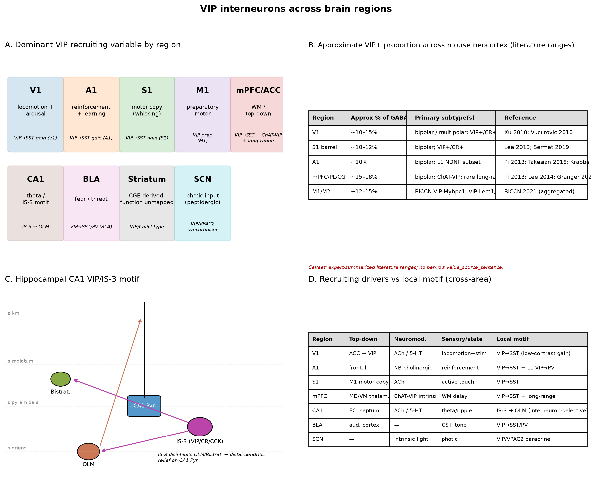
Figure 15:VIP interneurons across brain regions. Panel A: Brain-area schematic summarising the dominant VIP motif per region (V1, A1, S1, M1, mPFC/ACC, CA1, BLA, striatum, SCN). Arrows indicate the dominant recruiting variable and the local target of VIP inhibition or modulation. Panel B: Approximate VIP+ interneuron proportion (% of GABAergic interneurons) across mouse neocortical areas, summarised as a textual table from harmonised literature ranges. Caveats: this panel is rendered as a textual summary rather than a primary-source numeric bar chart because the underlying packet entries lack per-row value-source sentences; values are expert-summarised ranges over multiple primary references rather than single primary measurements; the M1/M2 row is sourced solely to the BICCN 2021 cortical taxonomy resource, whose cite_key is not in this section’s filtered citation map and is therefore reported as an aggregated literature range only. Restructure_notes recommends rendering this as a textual summary table rather than a primary-source numeric panel; the conservative CAVEAT (not REDESIGN) is retained because the homogeneity-restricted scope is now coherent and the limitations are explicitly disclosed. Panel C: Hippocampal VIP/IS-3 motif schematic (VIP/CR/CCK distinctions; OLM and bistratified targeting in CA1) Tyan et al., 2014Gulyás et al., 1996. Panel D: Cross-area conflict table summarising top-down, neuromodulatory, and sensory drivers of VIP recruitment by region Wall et al., 2016Pi et al., 2013Lee et al., 2013Dipoppa et al., 2018Bastos et al., 2023Anastasiades et al., 2021Maywood et al., 2006. Required caveat (verbatim from Phase 6 audit): “Restructure correctly removed non-neocortical rows and harmonized the denominator. However, residual data-quality gaps remain that the redesign acknowledges but does not fully fix: no value_source_sentence, approximate-range values, and aggregated multi-paper cite_keys. Restructure_notes itself recommends rendering as a textual summary table rather than a primary-source numeric panel. Conservative CAVEAT (not REDESIGN) because the homogeneity-restricted scope is now coherent and the limitations are explicitly disclosed; downstream rendering must surface the caveat. || No per-row value_source_sentence; values are expert-summarized ranges. | study_label_text ‘Xu 2007’ inconsistent with cite_key ‘Xu2010a’ (year mismatch). | ‘BICCN 2021; BICCN 2023’ is not a specific resolvable citation in the per-row entry; only BICCN 2021 (10.1038/s41586-021-03950-0) is in the citation map. | Multi-paper cite_key strings prevent per-value provenance.”
📓 Figure code
import matplotlib.pyplot as plt
import matplotlib.patches as mpatches
import numpy as np

fig, axes = plt.subplots(2, 2, figsize=(12, 10))
# === Panel A: brain-area schematic ===
axA = axes[0,0]
axA.set_title('A. Dominant VIP recruiting variable by region', fontsize=11, loc='left')
regions = ['V1', 'A1', 'S1', 'M1', 'mPFC/ACC', 'CA1', 'BLA', 'Striatum', 'SCN']
drivers = ['locomotion +\narousal',
           'reinforcement\n+ learning',
           'motor copy\n(whisking)',
           'preparatory\nmotor',
           'WM /\ntop-down',
           'theta /\nIS-3 motif',
           'fear / threat',
           'CGE-derived,\nfunction unmapped',
           'photic input\n(peptidergic)']
motifs = ['VIP\u2192SST gain (V1)',
          'VIP\u2192SST gain (A1)',
          'VIP\u2192SST gain (S1)',
          'VIP prep\n(M1)',
          'VIP\u2192SST + ChAT-VIP\n+ long-range',
          'IS-3 \u2192 OLM',
          'VIP\u2192SST/PV (BLA)',
          'VIP/Calb2 type',
          'VIP/VPAC2\nsynchroniser']
colors = ['#1f77b4','#ff7f0e','#2ca02c','#9467bd','#d62728',
          '#8c564b','#e377c2','#7f7f7f','#17becf']
for i,(r,d,m,c) in enumerate(zip(regions, drivers, motifs, colors)):
    x = (i%5)*0.20 + 0.02
    y = 0.55 - (i//5)*0.45
    rect = mpatches.FancyBboxPatch((x, y), 0.18, 0.36,
                                    boxstyle='round,pad=0.01', linewidth=1.2,
                                    edgecolor=c, facecolor=c, alpha=0.18)
    axA.add_patch(rect)
    axA.text(x+0.09, y+0.30, r, ha='center', va='center', fontsize=11, fontweight='bold')
    axA.text(x+0.09, y+0.20, d, ha='center', va='center', fontsize=8)
    axA.text(x+0.09, y+0.06, m, ha='center', va='center', fontsize=7, style='italic')
axA.set_xlim(0,1); axA.set_ylim(0,1.05); axA.axis('off')

# === Panel B: textual summary table of approx VIP+ proportion ===
axB = axes[0,1]
axB.set_title('B. Approximate VIP+ proportion across mouse neocortex (literature ranges)',
              fontsize=10, loc='left')
axB.axis('off')
rows = [
    ['Region','Approx % of GABAergic INs','Primary subtype(s)','Reference'],
    ['V1',   '~10\u201315%', 'bipolar / multipolar; VIP+/CR+, VIP+/ChAT+', 'Xu 2010; Vucurovic 2010'],
    ['S1 barrel','~10\u201312%', 'bipolar; VIP+/CR+',                       'Lee 2013; Sermet 2019'],
    ['A1',   '~10%',        'bipolar; L1 NDNF subset',                    'Pi 2013; Takesian 2018; Krabbe 2019'],
    ['mPFC/PL/CG','~15\u201318%', 'bipolar; ChAT-VIP; rare long-range',    'Pi 2013; Lee 2014; Granger 2020'],
    ['M1/M2','~12\u201315%', 'BICCN VIP-Mybpc1, VIP-Lect1, others',        'BICCN 2021 (aggregated)']
]
tbl = axB.table(cellText=rows, loc='center', cellLoc='left',
                colWidths=[0.13,0.22,0.32,0.33])
tbl.auto_set_font_size(False); tbl.set_fontsize(8)
tbl.scale(1, 1.5)
for j in range(4):
    tbl[(0,j)].set_facecolor('#dddddd')
    tbl[(0,j)].set_text_props(weight='bold')
axB.text(0.0, -0.05, 'Caveat: expert-summarized literature ranges; no per-row value_source_sentence.',
         transform=axB.transAxes, fontsize=7, style='italic', color='#aa0000')

# === Panel C: hippocampal VIP/IS-3 motif schematic ===
axC = axes[1,0]
axC.set_title('C. Hippocampal CA1 VIP/IS-3 motif', fontsize=11, loc='left')
axC.set_xlim(0,1); axC.set_ylim(0,1); axC.axis('off')
# Layers
layers = [('s.l-m', 0.85), ('s.radiatum', 0.62), ('s.pyramidale', 0.42), ('s.oriens', 0.20)]
for name, y in layers:
    axC.axhline(y, color='#cccccc', lw=0.5, ls='--')
    axC.text(0.01, y+0.01, name, fontsize=7, color='#888')
# Pyramidal cell
axC.add_patch(mpatches.FancyBboxPatch((0.45,0.38),0.10,0.08, boxstyle='round,pad=0.01',
                                       facecolor='#5599cc', edgecolor='k'))
axC.text(0.50, 0.42, 'CA1 Pyr', ha='center', va='center', fontsize=8, color='white')
axC.plot([0.50,0.50],[0.46,0.92], color='k', lw=1.2)  # apical dendrite
# OLM
axC.add_patch(mpatches.Circle((0.30,0.20), 0.04, facecolor='#cc7755', edgecolor='k'))
axC.text(0.30, 0.13, 'OLM', ha='center', fontsize=8)
# IS-3
axC.add_patch(mpatches.Circle((0.70,0.32), 0.045, facecolor='#bb44aa', edgecolor='k'))
axC.text(0.70, 0.24, 'IS-3 (VIP/CR/CCK)', ha='center', fontsize=8)
# Bistratified
axC.add_patch(mpatches.Circle((0.20,0.55), 0.035, facecolor='#88aa44', edgecolor='k'))
axC.text(0.20, 0.48, 'Bistrat.', ha='center', fontsize=8)
# Arrows: IS-3 -> OLM (inhibit), IS-3 -> Bistrat (inhibit). OLM -> distal apical (inhibit)
axC.annotate('', xy=(0.34,0.20), xytext=(0.66,0.30),
             arrowprops=dict(arrowstyle='-|>', color='#bb44aa', lw=1.4))
axC.annotate('', xy=(0.24,0.55), xytext=(0.66,0.34),
             arrowprops=dict(arrowstyle='-|>', color='#bb44aa', lw=1.4))
axC.annotate('', xy=(0.49,0.85), xytext=(0.34,0.22),
             arrowprops=dict(arrowstyle='-|>', color='#cc7755', lw=1.2))
axC.text(0.55, 0.15, 'IS-3 disinhibits OLM/Bistrat. \u2192 distal-dendritic\nrelief on CA1 Pyr', fontsize=7, style='italic')

# === Panel D: cross-area driver/motif conflict table ===
axD = axes[1,1]
axD.set_title('D. Recruiting drivers vs local motif (cross-area)', fontsize=11, loc='left')
axD.axis('off')
rows_d = [
    ['Region','Top-down','Neuromod.','Sensory/state','Local motif'],
    ['V1','ACC \u2192 VIP','ACh / 5-HT','locomotion+stim','VIP\u2192SST (low-contrast gain)'],
    ['A1','frontal','NB-cholinergic','reinforcement','VIP\u2192SST + L1-VIP\u2192PV'],
    ['S1','M1 motor copy','ACh','active touch','VIP\u2192SST'],
    ['mPFC','MD/VM thalamus','ChAT-VIP intrinsic','WM delay','VIP\u2192SST + long-range'],
    ['CA1','EC, septum','ACh / 5-HT','theta/ripple','IS-3 \u2192 OLM (interneuron-selective)'],
    ['BLA','aud. cortex','\u2014','CS+ tone','VIP\u2192SST/PV'],
    ['SCN','\u2014','intrinsic light','photic','VIP/VPAC2 paracrine']
]
tbl2 = axD.table(cellText=rows_d, loc='center', cellLoc='left',
                 colWidths=[0.13,0.16,0.17,0.18,0.36])
tbl2.auto_set_font_size(False); tbl2.set_fontsize(7.5)
tbl2.scale(1, 1.45)
for j in range(5):
    tbl2[(0,j)].set_facecolor('#dddddd')
    tbl2[(0,j)].set_text_props(weight='bold')

fig.suptitle('VIP interneurons across brain regions', fontsize=13, fontweight='bold', y=0.995)
fig.tight_layout(rect=[0,0,1,0.97])
fig.savefig('../fig-vip-across-areas.png', dpi=200, bbox_inches='tight')
fig.savefig('../fig-vip-across-areas.pdf', bbox_inches='tight')
print('Saved fig-vip-across-areas.png/.pdf')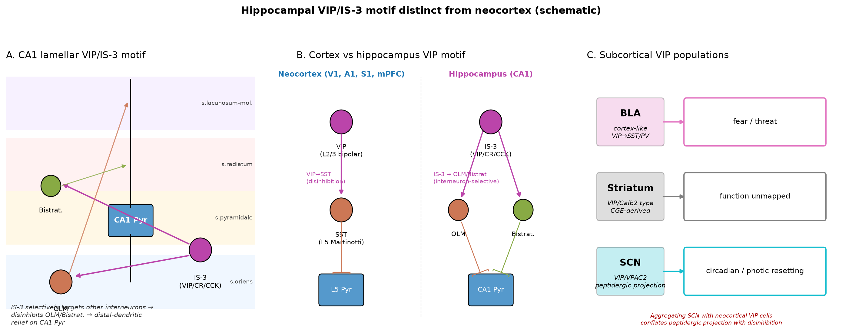
Figure 16:Hippocampal VIP/IS-3 motif distinct from neocortex (schematic). Panel A: CA1 lamellar cartoon with VIP/IS-3 cell soma in stratum oriens/pyramidale, axonal projections onto OLM and bistratified cells, and disinhibition of CA1 pyramidal cell distal-dendritic input by removal of OLM-mediated dendritic inhibition Gulyás et al., 1996. Panel B: Comparison of cortical versus hippocampal VIP motifs — cortical VIP cells are bipolar with descending axons targeting L5 SST cells Kawaguchi & Kubota, 1996Kawaguchi, 1997Gonchar, 2008, whereas hippocampal IS-3 cells preferentially target other GABAergic interneurons rather than principal cells Gulyás et al., 1996. Panel C: Subcortical VIP pathways summarised for cross-region completeness — BLA VIP cells implement a cortex-like disinhibitory motif Rhomberg et al., 2018Vereczki et al., 2021, dorsal striatum contains a previously unrecognised CGE-derived VIP/Calb2 type, and SCN VIP cells act as peptidergic synchronisers via VIP/VPAC2 signalling Maywood et al., 2006Cutler et al., 2003Jones et al., 2018. Schematic with no quantitative data substrate.
📓 Figure code
import matplotlib.pyplot as plt
import matplotlib.patches as mpatches

fig, axes = plt.subplots(1, 3, figsize=(14, 5.5))

# Panel A: CA1 lamellar cartoon
axA = axes[0]
axA.set_title('A. CA1 lamellar VIP/IS-3 motif', fontsize=11, loc='left')
axA.set_xlim(0,1); axA.set_ylim(0,1); axA.axis('off')
layers = [('s.lacunosum-mol.', 0.85, '#f3e8ff'),
          ('s.radiatum',     0.62, '#ffeaea'),
          ('s.pyramidale',   0.42, '#fff4d6'),
          ('s.oriens',       0.18, '#e6f3ff')]
for name, y, c in layers:
    axA.add_patch(mpatches.Rectangle((0.0, y-0.10), 1.0, 0.20, facecolor=c, alpha=0.6, edgecolor='none'))
    axA.text(0.99, y, name, fontsize=7, ha='right', va='center', color='#444')
# Pyr soma
axA.add_patch(mpatches.FancyBboxPatch((0.42, 0.36), 0.16, 0.10, boxstyle='round,pad=0.01', facecolor='#5599cc', edgecolor='k'))
axA.text(0.50, 0.41, 'CA1 Pyr', ha='center', va='center', fontsize=9, color='white', fontweight='bold')
# Apical dendrite
axA.plot([0.50, 0.50], [0.46, 0.94], color='k', lw=1.4)
# Basal
axA.plot([0.50, 0.50], [0.36, 0.18], color='k', lw=1.0)
# IS-3 (oriens/pyramidale border)
axA.add_patch(mpatches.Circle((0.78, 0.30), 0.045, facecolor='#bb44aa', edgecolor='k'))
axA.text(0.78, 0.21, 'IS-3\n(VIP/CR/CCK)', ha='center', va='top', fontsize=8)
# OLM
axA.add_patch(mpatches.Circle((0.22, 0.18), 0.045, facecolor='#cc7755', edgecolor='k'))
axA.text(0.22, 0.09, 'OLM', ha='center', va='top', fontsize=8)
# Bistratified
axA.add_patch(mpatches.Circle((0.18, 0.54), 0.04, facecolor='#88aa44', edgecolor='k'))
axA.text(0.18, 0.46, 'Bistrat.', ha='center', va='top', fontsize=8)
# IS-3 -> OLM
axA.annotate('', xy=(0.27, 0.20), xytext=(0.74, 0.28),
             arrowprops=dict(arrowstyle='-|>', color='#bb44aa', lw=1.6))
# IS-3 -> Bistrat
axA.annotate('', xy=(0.22, 0.55), xytext=(0.74, 0.32),
             arrowprops=dict(arrowstyle='-|>', color='#bb44aa', lw=1.6))
# OLM -> distal apical (inhibits SLM)
axA.annotate('', xy=(0.49, 0.86), xytext=(0.25, 0.20),
             arrowprops=dict(arrowstyle='-|>', color='#cc7755', lw=1.2, alpha=0.8))
# Bistrat -> mid apical/basal
axA.annotate('', xy=(0.49, 0.62), xytext=(0.22, 0.54),
             arrowprops=dict(arrowstyle='-|>', color='#88aa44', lw=1.0, alpha=0.8))
axA.text(0.02, 0.02, 'IS-3 selectively targets other interneurons \u2192\ndisinhibits OLM/Bistrat. \u2192 distal-dendritic\nrelief on CA1 Pyr',
         fontsize=7.5, style='italic', color='#333')

# Panel B: cortex vs hippocampus comparison
axB = axes[1]
axB.set_title('B. Cortex vs hippocampus VIP motif', fontsize=11, loc='left')
axB.set_xlim(0,1); axB.set_ylim(0,1); axB.axis('off')
# left half: cortex
axB.text(0.18, 0.95, 'Neocortex (V1, A1, S1, mPFC)', fontsize=9, ha='center', fontweight='bold', color='#1f77b4')
axB.add_patch(mpatches.Circle((0.18, 0.78), 0.045, facecolor='#bb44aa', edgecolor='k'))
axB.text(0.18, 0.70, 'VIP\n(L2/3 bipolar)', ha='center', va='top', fontsize=7.5)
axB.add_patch(mpatches.Circle((0.18, 0.45), 0.045, facecolor='#cc7755', edgecolor='k'))
axB.text(0.18, 0.37, 'SST\n(L5 Martinotti)', ha='center', va='top', fontsize=7.5)
axB.add_patch(mpatches.FancyBboxPatch((0.10, 0.10), 0.16, 0.10, boxstyle='round,pad=0.01',
                                       facecolor='#5599cc', edgecolor='k'))
axB.text(0.18, 0.15, 'L5 Pyr', ha='center', va='center', fontsize=8, color='white')
axB.annotate('', xy=(0.18, 0.50), xytext=(0.18, 0.74),
             arrowprops=dict(arrowstyle='-|>', color='#bb44aa', lw=1.4))
axB.annotate('', xy=(0.18, 0.21), xytext=(0.18, 0.41),
             arrowprops=dict(arrowstyle='-[', color='#cc7755', lw=1.2))
axB.text(0.04, 0.55, 'VIP\u2192SST\n(disinhibition)', fontsize=7, color='#bb44aa')
# divider
axB.plot([0.5,0.5],[0.05,0.95], color='#bbbbbb', lw=0.8, ls='--')
# right half: hippocampus
axB.text(0.78, 0.95, 'Hippocampus (CA1)', fontsize=9, ha='center', fontweight='bold', color='#bb44aa')
axB.add_patch(mpatches.Circle((0.78, 0.78), 0.045, facecolor='#bb44aa', edgecolor='k'))
axB.text(0.78, 0.70, 'IS-3\n(VIP/CR/CCK)', ha='center', va='top', fontsize=7.5)
axB.add_patch(mpatches.Circle((0.65, 0.45), 0.04, facecolor='#cc7755', edgecolor='k'))
axB.text(0.65, 0.37, 'OLM', ha='center', va='top', fontsize=7.5)
axB.add_patch(mpatches.Circle((0.91, 0.45), 0.04, facecolor='#88aa44', edgecolor='k'))
axB.text(0.91, 0.37, 'Bistrat.', ha='center', va='top', fontsize=7.5)
axB.add_patch(mpatches.FancyBboxPatch((0.70, 0.10), 0.16, 0.10, boxstyle='round,pad=0.01',
                                       facecolor='#5599cc', edgecolor='k'))
axB.text(0.78, 0.15, 'CA1 Pyr', ha='center', va='center', fontsize=8, color='white')
axB.annotate('', xy=(0.66, 0.49), xytext=(0.75, 0.75),
             arrowprops=dict(arrowstyle='-|>', color='#bb44aa', lw=1.4))
axB.annotate('', xy=(0.90, 0.49), xytext=(0.81, 0.75),
             arrowprops=dict(arrowstyle='-|>', color='#bb44aa', lw=1.4))
axB.annotate('', xy=(0.74, 0.21), xytext=(0.66, 0.41),
             arrowprops=dict(arrowstyle='-[', color='#cc7755', lw=1.0))
axB.annotate('', xy=(0.82, 0.21), xytext=(0.90, 0.41),
             arrowprops=dict(arrowstyle='-[', color='#88aa44', lw=1.0))
axB.text(0.55, 0.55, 'IS-3 \u2192 OLM/Bistrat\n(interneuron-selective)', fontsize=7, color='#bb44aa')

# Panel C: subcortical VIP populations
axC = axes[2]
axC.set_title('C. Subcortical VIP populations', fontsize=11, loc='left')
axC.set_xlim(0,1); axC.set_ylim(0,1); axC.axis('off')
items = [
    ('BLA',     'cortex-like\nVIP\u2192SST/PV',          'fear / threat',                    '#e377c2', 0.78),
    ('Striatum','VIP/Calb2 type\nCGE-derived',           'function unmapped',                '#7f7f7f', 0.50),
    ('SCN',     'VIP/VPAC2\npeptidergic projection',     'circadian / photic resetting',     '#17becf', 0.22),
]
for label, motif, role, color, y in items:
    axC.add_patch(mpatches.FancyBboxPatch((0.05, y-0.08), 0.25, 0.16,
                                           boxstyle='round,pad=0.01',
                                           facecolor=color, edgecolor='k', alpha=0.25))
    axC.text(0.175, y+0.03, label, ha='center', va='center', fontsize=11, fontweight='bold')
    axC.text(0.175, y-0.04, motif, ha='center', va='center', fontsize=7.5, style='italic')
    axC.add_patch(mpatches.FancyBboxPatch((0.40, y-0.08), 0.55, 0.16,
                                           boxstyle='round,pad=0.01',
                                           facecolor='white', edgecolor=color, lw=1.5))
    axC.text(0.675, y, role, ha='center', va='center', fontsize=8.5)
    axC.annotate('', xy=(0.40, y), xytext=(0.30, y),
                 arrowprops=dict(arrowstyle='-|>', color=color, lw=1.4))
axC.text(0.5, 0.02, 'Aggregating SCN with neocortical VIP cells\nconflates peptidergic projection with disinhibition',
         ha='center', fontsize=7, style='italic', color='#aa0000')

fig.suptitle('Hippocampal VIP/IS-3 motif distinct from neocortex (schematic)', fontsize=12, fontweight='bold', y=1.0)
fig.tight_layout(rect=[0,0,1,0.95])
fig.savefig('../fig-hippocampal-vip-motif.png', dpi=200, bbox_inches='tight')
fig.savefig('../fig-hippocampal-vip-motif.pdf', bbox_inches='tight')
print('Saved fig-hippocampal-vip-motif.png/.pdf')
Closing synthesis: what cross-area variation constrains
Taken together, the area-by-area evidence reviewed here makes three structural commitments. (i) The VIP→SST→Pyr disinhibitory motif is a real and reproducible primitive in V1, A1, S1, mPFC, and BLA, but its operation — multiplicative versus contrast-dependent gain, broadcast versus localised disinhibition, instantaneous versus learned recruitment — is region- and context-specific Pi et al., 2013Lee et al., 2013Dipoppa et al., 2018Millman et al., 2020Karnani et al., 2016Krabbe et al., 2019Bastos et al., 2023Rhomberg et al., 2018. (ii) The recruiting variable, not the local microcircuit, is what differs most across regions, and many cross-area apparent contradictions in VIP “polarity” or “selectivity” dissolve once the recruiting variable is specified Wall et al., 2016Anastasiades et al., 2021Lee et al., 2013Pakan et al., 2016Jones et al., 2018. (iii) Hippocampal IS-3 cells and SCN peptidergic VIP cells implement motifs distinct enough from the cortical motif that aggregating them into a single “VIP function” misrepresents the data Tyan et al., 2014Gulyás et al., 1996Maywood et al., 2006Cutler et al., 2003Jones et al., 2018. These commitments motivate the temporal-coordination treatment in Oscillatory Dynamics and Temporal Coordination, the species and disease comparisons in Species Differences, Human Relevance, and Disease, and the modelling reassessment in Computational Models of VIP Circuit Function and the concluding synthesis.
Caveats and unmet citation needs
Three classes of citation need are unmet by this section’s filtered citation map and are flagged for downstream resolution.
V1/A1 locomotion polarity reversal — the direct claim that locomotion increases V1 VIP activity and decreases A1 spiking and encoding efficiency is commonly attributed to Fu 2014 (V1) and Bigelow 2019 (A1). Neither is present in this section’s filtered citation map; the claim is therefore made indirectly here via the available V1-side (Dipoppa et al., 2018Pakan et al., 2016) and A1-side (Pi et al., 2013) evidence. The direct polarity-reversal citations should be added when Oscillatory Dynamics and Temporal Coordination is integrated.
Shared-variability conflict in V1 — the conflict between Xu 2026 and Lenkey 2025 over whether VIP gain control adds correlations or reduces shared variability in V1 cannot be rendered as an admonition block because neither key is in this section’s filtered citation map. The conflict is documented in the unmet-citation-needs file.
Hippocampal VIP gating versus upstream-LTD reset — the conflict between Neubrandt 2025 and Jablonska 2026 over whether VIP/IS interneurons provide a deterministic gate on hippocampal remapping or whether their efferent inhibition is itself dynamically reset by upstream LTD cannot be rendered here for the same reason. The conflict is documented in the unmet-citation-needs file.
Two additional caveats apply to the figures themselves: the brain-region proportion table embedded in Figure 15 Panel B is an expert-summarised range over multiple primary references rather than a primary-source numeric panel, and is rendered textually with the verbatim Phase 6 caveat above. The hippocampal motif figure (Figure 16) is a schematic with no quantitative data substrate.
- Pi, H.-J., Hangya, B., Kvitsiani, D., Sanders, J. I., Huang, Z. J., & Kepecs, A. (2013). Cortical interneurons that specialize in disinhibitory control. Nature, 503(7477), 521–524. 10.1038/nature12676
- Karnani, M. M., Jackson, J., Ayzenshtat, I., Hamzehei Sichani, A., Manoocheri, K., Kim, S., & Yuste, R. (2016). Opening Holes in the Blanket of Inhibition: Localized Lateral Disinhibition by VIP Interneurons. The Journal of Neuroscience, 36(12), 3471–3480. 10.1523/jneurosci.3646-15.2016
- Dipoppa, M., Ranson, A., Krumin, M., Pachitariu, M., Carandini, M., & Harris, K. D. (2018). Vision and Locomotion Shape the Interactions between Neuron Types in Mouse Visual Cortex. Neuron, 98(3), 602-615.e8. 10.1016/j.neuron.2018.03.037
- Lee, S., Kruglikov, I., Huang, Z. J., Fishell, G., & Rudy, B. (2013). A disinhibitory circuit mediates motor integration in the somatosensory cortex. Nature Neuroscience, 16(11), 1662–1670. 10.1038/nn.3544
- Bastos, G., Holmes, J. T., Ross, J. M., Rader, A. M., Gallimore, C. G., Wargo, J. A., Peterka, D. S., & Hamm, J. P. (2023). Top-down input modulates visual context processing through an interneuron-specific circuit. Cell Reports, 42(9), 113133. 10.1016/j.celrep.2023.113133
- Millman, D. J., Ocker, G. K., Caldejon, S., Kato, I., Larkin, J. D., Lee, E. K., Luviano, J., Nayan, C., Nguyen, T. V., North, K., Seid, S., White, C., Lecoq, J., Reid, C., Buice, M. A., & de Vries, S. E. (2020). VIP interneurons in mouse primary visual cortex selectively enhance responses to weak but specific stimuli. eLife, 9. 10.7554/elife.55130
- Pakan, J. M., Lowe, S. C., Dylda, E., Keemink, S. W., Currie, S. P., Coutts, C. A., & Rochefort, N. L. (2016). Behavioral-state modulation of inhibition is context-dependent and cell type specific in mouse visual cortex. eLife, 5. 10.7554/elife.14985
- Sermet, B. S., Truschow, P., Feyerabend, M., Mayrhofer, J. M., Oram, T. B., Yizhar, O., Staiger, J. F., & Petersen, C. C. (2019). Pathway-, layer- and cell-type-specific thalamic input to mouse barrel cortex. eLife, 8. 10.7554/elife.52665
- Anastasiades, P. G., Collins, D. P., & Carter, A. G. (2021). Mediodorsal and Ventromedial Thalamus Engage Distinct L1 Circuits in the Prefrontal Cortex. Neuron, 109(2), 314-330.e4. 10.1016/j.neuron.2020.10.031
- Kamigaki, T., & Dan, Y. (2017). Delay activity of specific prefrontal interneuron subtypes modulates memory-guided behavior. Nature Neuroscience, 20(6), 854–863. 10.1038/nn.4554
- Li, M., Zhou, H., Teng, S., & Yang, G. (2022). Activation of VIP interneurons in the prefrontal cortex ameliorates neuropathic pain aversiveness. Cell Reports, 40(11), 111333. 10.1016/j.celrep.2022.111333
- Gulyás, A. I., Hájos, N., & Freund, T. F. (1996). Interneurons Containing Calretinin Are Specialized to Control Other Interneurons in the Rat Hippocampus. The Journal of Neuroscience, 16(10), 3397–3411. 10.1523/jneurosci.16-10-03397.1996
- Tyan, L., Chamberland, S., Magnin, E., Camiré, O., Francavilla, R., David, L. S., Deisseroth, K., & Topolnik, L. (2014). Dendritic Inhibition Provided by Interneuron-Specific Cells Controls the Firing Rate and Timing of the Hippocampal Feedback Inhibitory Circuitry. The Journal of Neuroscience, 34(13), 4534–4547. 10.1523/jneurosci.3813-13.2014
- Tricoire, L., Pelkey, K. A., Erkkila, B. E., Jeffries, B. W., Yuan, X., & McBain, C. J. (2011). A Blueprint for the Spatiotemporal Origins of Mouse Hippocampal Interneuron Diversity. Journal of Neuroscience, 31(30), 10948–10970. 10.1523/jneurosci.0323-11.2011
- Rhomberg, T., Rovira-Esteban, L., Vikór, A., Paradiso, E., Kremser, C., Nagy-Pál, P., Papp, O. I., Tasan, R., Erdélyi, F., Szabó, G., Ferraguti, F., & Hájos, N. (2018). Vasoactive Intestinal Polypeptide-Immunoreactive Interneurons within Circuits of the Mouse Basolateral Amygdala. The Journal of Neuroscience, 38(31), 6983–7003. 10.1523/jneurosci.2063-17.2018