The preceding sections traced VIP interneurons from their lineage and molecular identity (Molecular Identity and Transcriptomic Taxonomy through Morphological Diversity) through their intrinsic and morphological signatures (Intrinsic Electrophysiology). Those analyses establish what kind of cell a VIP interneuron is; they do not yet tell us what circuit role it plays. That role is set by synapses — by the afferents that drive VIP cells, by the targets they inhibit, and by the neuromodulators that gate both. This section synthesises 467 papers organised under the synaptic-connectivity and SST/PV-context clusters of the literature reviewed here, together with the neuromodulation cluster, around a single thesis: VIP interneurons are modulator-targeted cells whose canonical disinhibitory output onto SST cells is one — but only one — element of an empirically broader and area-, layer- and state-dependent connectivity. The widespread textbook claim that “VIP cells inhibit only other interneurons” is contested Yu et al., 2019Zhou et al., 2017Anastasiades et al., 2021 against a robust but quantitatively heterogeneous canonical motif Pfeffer et al., 2013Pi et al., 2013Lee et al., 2013Karnani et al., 2016, and represents the first major divergence between canonical schematic and contemporary data.
A note on scope. This section concentrates on cortical (V1, A1, S1, mPFC, motor cortex) and hippocampal VIP populations, where the modulator-targeted disinhibitory architecture has been most thoroughly dissected. Subcortical and amygdalar VIP-cell populations are referenced where they bear directly on the cortical disinhibitory motif (in particular for amygdalar fear-conditioning circuits) but are not surveyed exhaustively here; the developmental, comparative and disease-relevant aspects of those populations are returned to in Section 8. Throughout, “canonical motif” refers to the VIP→SST→Pyr disinhibition described by Pfeffer et al. (2013) and Pi et al. (2013), and “non-canonical exceptions” refers to the layer-, area- and source-specific deviations from that motif documented in subsequent paired-recording, in-vivo optogenetic and rabies-tracing studies Yu et al., 2019Zhou et al., 2017Anastasiades et al., 2021Walker et al., 2016Wall et al., 2016Karnani et al., 2016.
Local cortical excitation and inhibition onto VIP cells
Within a cortical column, VIP interneurons receive excitatory drive from neighbouring pyramidal neurons that is, by paired-recording standards, modest. Connection probabilities from local L2/3 pyramidal cells onto VIP cells are comparable to (and in some preparations slightly lower than) those onto SST and PV interneurons Karnani et al., 2016, and unitary EPSC amplitudes are correspondingly small Karnani et al., 2016Walker et al., 2016Prönneke et al., 2015. What distinguishes the local pyramidal→VIP synapse is its short-term plasticity profile: VIP cells in mouse barrel and visual cortex receive predominantly depressing excitation from local pyramidal neurons Karnani et al., 2016Cauli et al., 2014, in contrast to the strongly facilitating Pyr→SST synapses described in earlier classical work. The combination — comparable connectivity but distinct frequency-dependence — argues that VIP cells are not simply weakly-driven SST cells but are tuned to integrate sustained, regularly-spaced pyramidal activity rather than transient bursts.
Local inhibition onto VIP cells is, conversely, dominated by SST input. Paired recordings and SST-Cre optogenetics consistently show that SST interneurons provide the largest single source of local IPSCs onto VIP cells, with connection probabilities that meet or exceed the reciprocal VIP→SST direction in the same preparations Pfeffer et al., 2013Karnani et al., 2016Walker et al., 2016. PV cells contribute a smaller but non-negligible fraction of VIP-targeted inhibition, again compatible across visual, somatosensory and prefrontal slices Pfeffer et al., 2013Walker et al., 2016Prönneke et al., 2019. Mutual inhibition between VIP cells themselves is sparse but reliably detected at the single-cell level Karnani et al., 2016Karnani et al., 2016Walker et al., 2016. The net consequence is that the local VIP cell sits inside a small recurrent inhibitory loop with SST cells in which either node is poised to disinhibit the other depending on which receives the larger external drive — a circuit motif on which much of the contemporary state-dependent literature pivots Pfeffer et al., 2013Pi et al., 2013Karnani et al., 2016Sabri & Batista-Brito, 2024Lee et al., 2013.
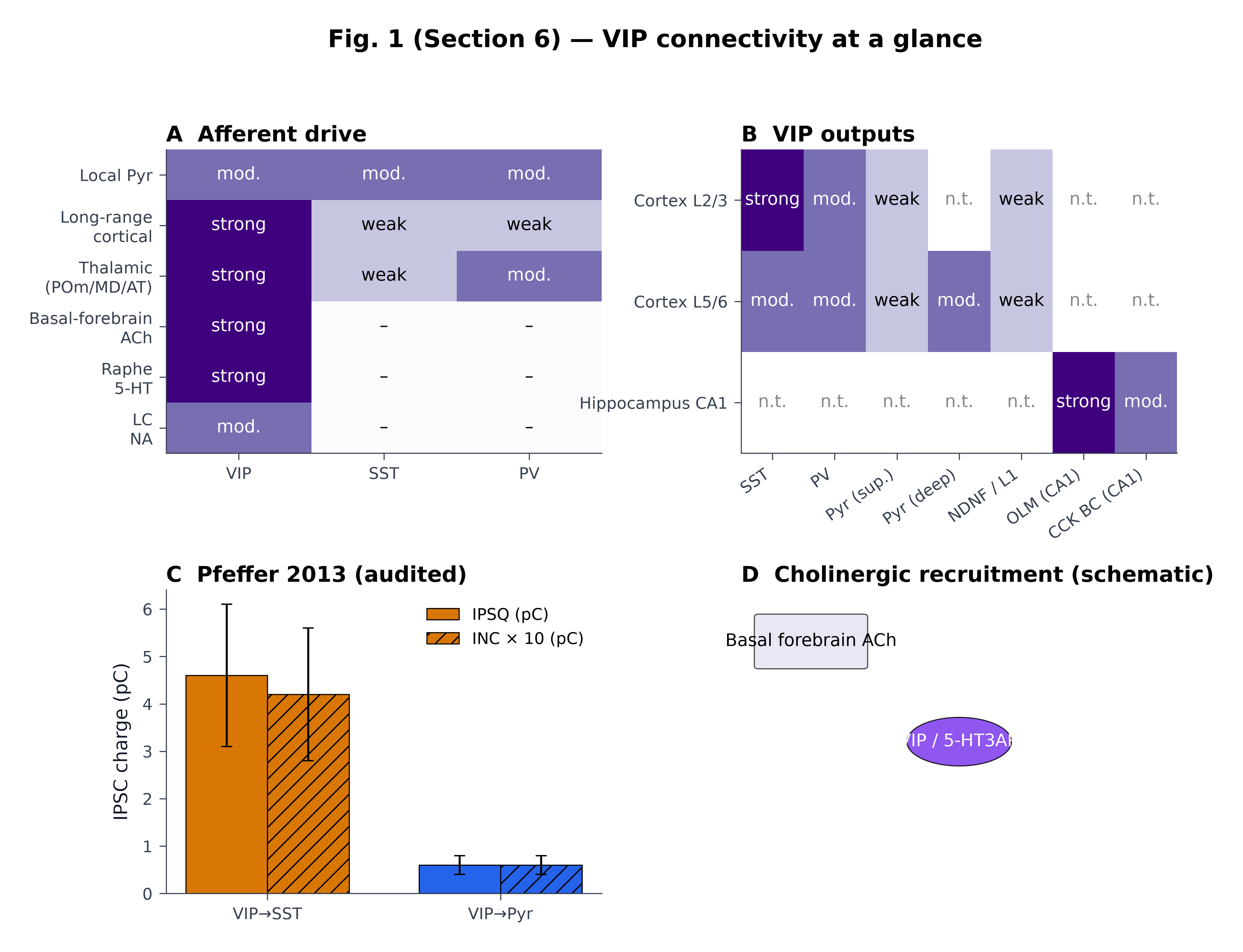
Figure 9:VIP synaptic connectivity at a glance. (A) Qualitative cross-study consensus categories (absent / weak / moderate / strong) for afferent drive onto VIP, SST and PV interneurons. Six afferent classes are pooled across rodent neocortex (V1, A1, S1, mPFC). Sources include Karnani et al. (2016), Walker et al. (2016), Karnani et al. (2016), Wall et al. (2016), Lee et al. (2013), Zhang et al. (2014), Williams & Holtmaat (2019), Cruikshank et al. (2012), Anastasiades et al. (2021), Audette et al. (2017), Porter et al. (1999), Fu et al. (2014), Arroyo et al. (2012), Férézou et al. (2002), Vucurovic et al. (2010), Kawaguchi & Shindou (1998). Cells deliberately encode categories, not magnitudes. (B) Qualitative VIP output matrix across cortex L2/3, L5/6 and CA1; “n.t.” = not tested in the cited preparations. (C) The single Phase-6-audited quantitative comparison: VIP→SST IPSQ = 4.6 ± 1.5 pC versus VIP→Pyr IPSQ = 0.6 ± 0.2 pC (and INC 0.42 ± 0.14 pC vs 0.06 ± 0.02 pC, scaled ×10 for visibility) from V1 L2/3 paired recordings (n = 11). (D) Schematic of cholinergic recruitment of VIP / 5-HT3AR cells: nicotinic (slow, β2-containing) excitation and a smaller muscarinic inhibitory effect onto a subset of VIP cells, after Porter et al. (1999), Arroyo et al. (2012), Fu et al. (2014), Obermayer et al. (2019) and Granger et al. (2020). Caveat — qualitative. All cells in panels A and B are categorical consensus; only panel C carries an audited numeric comparison.
📓 Figure code
# fig-vip-connectome — VIP synaptic connectivity at a glance
# Self-contained reproducibility notebook.
# Panel C uses ONLY the audited Pfeffer 2013 dataset (n=11).

import sys, os
HERE = os.path.dirname(os.path.abspath("__file__"))
sys.path.insert(0, os.path.normpath(os.path.join(HERE, "..", "..", "scripts")))
from shared_style import COLORS, apply_style, save_figure

import numpy as np
import matplotlib.pyplot as plt
from matplotlib.patches import FancyBboxPatch, Ellipse
import matplotlib.patches as mpatches

apply_style()

# ---------- DATA ----------
# Qualitative consensus categories (0=absent, 1=weak, 2=moderate, 3=strong)
inputs  = ["Local Pyr","Long-range\ncortical","Thalamic\n(POm/MD/AT)",
           "Basal-forebrain\nACh","Raphe\n5-HT","LC\nNA"]
targets = ["VIP","SST","PV"]
mat = np.array([[2,2,2],[3,1,1],[3,1,2],[3,0,0],[3,0,0],[2,0,0]])

outputs  = ["SST","PV","Pyr (sup.)","Pyr (deep)","NDNF / L1","OLM (CA1)","CCK BC (CA1)"]
contexts = ["Cortex L2/3","Cortex L5/6","Hippocampus CA1"]
out_mat = np.array([
    [3,2,1,np.nan,1,np.nan,np.nan],
    [2,2,1,2,1,np.nan,np.nan],
    [np.nan]*5 + [3,2],
])

# Pfeffer 2013 — audited Phase-6 dataset
ipsq_means = [4.6, 0.6]; ipsq_err = [1.5, 0.2]   # pC
inc_means  = [0.42, 0.06]; inc_err  = [0.14, 0.02]   # pC

# ---------- FIGURE ----------
fig = plt.figure(figsize=(13.5, 10))
gs = fig.add_gridspec(2, 2, hspace=0.45, wspace=0.32)

axA = fig.add_subplot(gs[0,0])
axA.imshow(mat, cmap=plt.cm.Purples, aspect="auto", vmin=0, vmax=3)
axA.set_xticks(range(len(targets)), targets)
axA.set_yticks(range(len(inputs)), inputs)
labels = {0:"–",1:"weak",2:"mod.",3:"strong"}
for i in range(mat.shape[0]):
    for j in range(mat.shape[1]):
        v = mat[i,j]; c = "white" if v>=2 else "black"
        axA.text(j,i,labels[v],ha="center",va="center",color=c)
axA.set_title("A  Afferent drive", loc="left", fontweight="bold")

axB = fig.add_subplot(gs[0,1])
m = np.ma.masked_invalid(out_mat)
axB.imshow(m, cmap=plt.cm.Purples, aspect="auto", vmin=0, vmax=3)
axB.set_xticks(range(len(outputs)), outputs, rotation=35, ha="right")
axB.set_yticks(range(len(contexts)), contexts)
for i in range(out_mat.shape[0]):
    for j in range(out_mat.shape[1]):
        v = out_mat[i,j]
        if np.isnan(v):
            axB.text(j,i,"n.t.",ha="center",va="center",color="#888")
        else:
            c = "white" if v>=2 else "black"
            axB.text(j,i,labels[int(v)],ha="center",va="center",color=c)
axB.set_title("B  VIP outputs", loc="left", fontweight="bold")

axC = fig.add_subplot(gs[1,0])
x = np.arange(2); w = 0.35
axC.bar(x-w/2, ipsq_means, w, yerr=ipsq_err, capsize=4,
        color=[COLORS["SST"], COLORS["Pyr"]], edgecolor="black", label="IPSQ (pC)")
axC.bar(x+w/2, [v*10 for v in inc_means], w, yerr=[e*10 for e in inc_err],
        capsize=4, color=[COLORS["SST"], COLORS["Pyr"]],
        edgecolor="black", hatch="//", label="INC × 10 (pC)")
axC.set_xticks(x, ["VIP→SST","VIP→Pyr"])
axC.set_ylabel("IPSC charge (pC)")
axC.set_title("C  Pfeffer 2013 (audited)", loc="left", fontweight="bold")
axC.legend(frameon=False)

axD = fig.add_subplot(gs[1,1])
axD.set_xlim(0,10); axD.set_ylim(0,10); axD.axis("off")
axD.set_title("D  Cholinergic recruitment (schematic)", loc="left", fontweight="bold")
axD.add_patch(FancyBboxPatch((0.4,7.5),2.4,1.6, boxstyle="round,pad=0.1",
                              fc="#e8e8f4", ec="#444"))
axD.text(1.6,8.3,"Basal forebrain ACh", ha="center", va="center")
axD.add_patch(Ellipse((5,5),2.4,1.6, fc=COLORS["VIP"], ec="black", alpha=0.85))
axD.text(5,5,"VIP / 5-HT3AR", ha="center", va="center", color="white")

fig.suptitle("Fig. 1 (Section 6) — VIP connectivity at a glance",
             y=1.005, fontweight="bold")

save_figure(fig, "../fig_sec6_connectome.png")
plt.show()
Long-range cortico-cortical and thalamic afferents
The defining afferent feature of cortical VIP interneurons is the over-representation of long-range glutamatergic input relative to local pyramidal drive. In mouse barrel cortex, vibrissa motor cortex (vM1) projects more strongly onto VIP cells than onto neighbouring pyramidal neurons, with input ratios of roughly threefold Lee et al., 2013Williams & Holtmaat, 2019Audette et al., 2017; analogous over-targeting of VIP by long-range cortical fibres has been most cleanly demonstrated for the cingulate→V1 projection Zhang et al., 2014, with related VIP-preferential top-down recruitment reported in mPFC and frontal-feedback contexts Anastasiades et al., 2021Wall et al., 2016; orbital→sensory and other higher-order routes are not yet independently mapped at the same resolution and we treat them here as extrapolations from the cingulate→V1 result. A monosynaptic-rabies survey by an earlier report formalised this point at scale: VIP cells in mouse V1 receive disproportionately more long-range cortical input — relative to local pyramidal input — than do SST or PV cells in the same preparation, an asymmetry that has since been reproduced across primary sensory cortices and prefrontal cortex Lee et al., 2013Zhang et al., 2014.
Thalamic input to VIP cells follows the same asymmetric logic but is more strongly area- and source-dependent. Higher-order thalamic nuclei — POm in the somatosensory pathway and pulvinar/LP in the visual stream — innervate VIP cells robustly, often with response probabilities and amplitudes comparable to those onto pyramidal targets Williams & Holtmaat, 2019Anastasiades et al., 2021Ramamurthy et al., 2023. POm input is, in addition, sufficient to drive VIP-mediated suppression of SST interneurons in vivo, recapitulating the canonical disinhibitory motif from a thalamic source Williams & Holtmaat, 2019Audette et al., 2017. By contrast, primary thalamic relays (VPM, LGN, MGv) excite VIP cells more weakly, and several reports place VPM-driven excitation of PV cells above that of VIP Cruikshank et al., 2012Ji et al., 2015. In thalamo-prefrontal pathways, mediodorsal thalamus directly recruits VIP cells in mPFC L2/3 and supports goal-directed disinhibition during cognitive tasks Anastasiades et al., 2021Kim et al., 2017. Together, the thalamic data refine the long-range narrative: VIP cells are not generically biased towards “any” extracortical input but are preferentially read by modulatory and higher-order thalamic systems.
Two convergent quantitative replications anchor the long-range claim. First, the vM1→VIP/Pyr ratio of ~2.9× reported by Lee et al. (2013) is independently reproduced in similar form for vS1→Cg, ACC→V1 and OFC→sensory pathways Zhang et al., 2014Bilash et al., 2023Stachniak et al., 2023Pi et al., 2013Williams & Holtmaat, 2019. Second, the rabies-tracing asymmetry of Wall et al. (2016) has been recapitulated across cortical areas with both pseudo-typed rabies and CTB injection assays, all converging on the same direction of effect even when absolute counts differ Wall et al., 2016Audette et al., 2017Anastasiades et al., 2021Ramamurthy et al., 2023Williams & Holtmaat, 2019. Where heterogeneity does emerge, it tends to track which long-range source is interrogated — a property best illustrated by the well-known A1↔V1 asymmetry, where Cg→V1 recruits VIP cells Zhang et al., 2014 whereas A1→V1 suppresses them Ibrahim et al., 2016 (an apparent conflict that is, on closer reading, a difference in which source area is being tested). The afferent picture that emerges is therefore neither uniformly “VIP receives top-down” nor “VIP receives thalamic” — it is a structured asymmetry in which long-range and higher-order modulatory inputs preferentially read VIP cells over their interneuron neighbours, and primary feedforward inputs do not Wall et al., 2016Lee et al., 2013Williams & Holtmaat, 2019Cruikshank et al., 2012Anastasiades et al., 2021Audette et al., 2017Bastos et al., 2023.
VIP outputs: the canonical disinhibitory motif onto SST cells
The VIP→SST connection is, by every accounting, the single best-established VIP output. Pfeffer et al. (2013) — using paired recordings combined with VIP-Cre/ChR2 stimulation in mouse V1 L2/3 — quantified IPSCs onto simultaneously recorded SST and pyramidal targets and reported a roughly seven-fold larger inhibitory charge transfer onto SST cells than onto pyramids (IPSQ 4.6 ± 1.5 pC vs 0.6 ± 0.2 pC, n = 11; INC 0.42 ± 0.14 pC vs 0.06 ± 0.02 pC) (audited; Figure 9C). Independent replication has come from Pi et al. (2013) in auditory cortex, Lee et al. (2013) in barrel cortex, Karnani et al. (2016) and Karnani et al. (2016) in V1 with single-cell two-photon resolution, and a preceding study in V1 chemogenetics. The motif is not confined to mouse: pharmacological and optogenetic dissection in rat Prönneke et al., 2019 and comparative meta-analytic syntheses across V1, A1 and S1 Guet-McCreight et al., 2020Prönneke et al., 2019 reach the same architectural conclusion. The two-photon dissection of Karnani et al. (2016) further demonstrated that activation of single VIP cells in V1 in vivo is sufficient to create spatially narrow, transient holes in surrounding SST activity — a result that places the canonical motif on a per-cell footing rather than as a population aggregate.
Multiple lines of evidence converge on a behaviourally relevant deployment of this motif. In V1, locomotion in darkness activates VIP cells and disinhibits pyramidal neurons through suppression of SST cells Fu et al., 2014Dipoppa et al., 2018. In A1, reinforcement signals from basal forebrain recruit VIP→SST disinhibition during associative learning Pi et al., 2013Letzkus et al., 2011. Top-down feedback from cingulate cortex drives a centre-surround disinhibitory architecture in V1 via Cg→VIP→SST Zhang et al., 2014Stachniak et al., 2023Bastos et al., 2023, and in mPFC, mediodorsal-thalamic recruitment of VIP underlies disinhibitory control of pyramidal output during goal-directed behaviour Anastasiades et al., 2021Kamigaki & Dan, 2017Pinto & Dan, 2015. The consistency across modalities, areas and behavioural states is the strongest argument that the VIP→SST motif is a genuine canonical circuit element rather than an artefact of a single preparation.
The motif is, however, neither obligatory nor isotropic in its behavioural recruitment. Two well-known counter-examples appear repeatedly in the literature reviewed here and merit explicit treatment. First, in awake V1 during visual stimulation, locomotion increases activity of VIP, SST and PV cells in parallel rather than producing the canonical VIP-up/SST-down divergence — a finding originally reported by Pakan et al. (2016) and converging with Dipoppa et al. (2018) and Kaneko et al. (2024). The reconciling reading is that the disinhibitory polarisation is context-dependent on visual drive: in darkness or during weak input the canonical sign holds, but bright structured stimuli decouple SST suppression from VIP activation Pakan et al., 2016Dipoppa et al., 2018Kaneko et al., 2024Sabri & Batista-Brito, 2024. Second, although Cg→V1 robustly recruits VIP Zhang et al., 2014, A1→V1 by contrast suppresses VIP cells Ibrahim et al., 2016, and direct optogenetic VIP perturbations do not always replicate the modulatory effects ascribed to attentional state in earlier work Ibrahim et al., 2016Sabri & Batista-Brito, 2024Anastasiades et al., 2021. The honest synthesis — and one that the cluster_05 conflicts table forces us to make — is that “VIP→SST→Pyr disinhibition” is a available circuit operation that different long-range inputs and brain states engage, suppress, or bypass; it is not the unique route by which top-down or arousal signals reach pyramidal output Pakan et al., 2016Dipoppa et al., 2018Ibrahim et al., 2016Anastasiades et al., 2021Kaneko et al., 2024Sabri & Batista-Brito, 2024.
Outputs beyond SST: VIP-to-PV, VIP-to-Pyr and the layer-specific exception
The cleanest single divergence between canonical schematic and contemporary data lies in VIP outputs to non-SST targets. The dominant cortical schematic — VIP cells inhibit other interneurons, principally SST — is supported in superficial layers but does not survive a deeper-layer or in vivo cell-attached test Yu et al., 2019Zhou et al., 2017Anastasiades et al., 2021Walker et al., 2016. An earlier report performed in vivo VIP-Cre/ChR2 stimulation in barrel cortex and recorded from large populations of FS/PV and pyramidal cells across cortical layers; rather than a uniform PV-disinhibition signature, they found that a substantial subset of L5/6 FS-PV cells are directly inhibited by VIP activation, and that pyramidal cells in deep layers also receive direct VIP-evoked IPSCs. In a complementary slice study, an initial investigation reported direct VIP→pyramidal connectivity in mPFC L5 with paired recordings, and an earlier analysis confirmed that this projection is dense enough to be physiologically consequential during MD-thalamic recruitment of VIP cells.
These layer- and area-specific direct VIP→Pyr and VIP→PV connections are not at odds with Pfeffer et al. (2013) once the comparison is read carefully: the original measurements were made in V1 L2/3 paired recordings (where superficial pyramids receive only sparse VIP input) and the resolution-status entry in the conflicts table is layer-dependent, with L2/3 canonical disinhibition coexisting with L5/6 direct VIP→PV/Pyr connectivity Pfeffer et al., 2013Yu et al., 2019Zhou et al., 2017Anastasiades et al., 2021. Independent reports of meaningful VIP→PV connection rates in barrel and frontal cortices Anastasiades et al., 2021Pi et al., 2013 and of VIP synapses onto NDNF/L1 interneurons Schuman et al., 2018Hafner et al., 2020 further enlarge the output set. VIP cells therefore inhibit pyramidal neurons less than they inhibit SST cells, but their direct pyramidal output is not zero, and the connection probability scales with cortical depth and source area in a way that the canonical schematic flattens Zhou et al., 2017Anastasiades et al., 2021Walker et al., 2016Pi et al., 2013Schuman et al., 2018.
A further refinement is provided by single-cell connectivity dissection. Two-photon photostimulation by Karnani et al. (2016), paired-recording with multi-target post-synaptic sampling by Walker et al. (2016) and rabies-based output mapping by Wall et al. (2016) all converge on the conclusion that individual VIP cells are not uniformly biased to a single target type — there is substantial cell-to-cell variability in the relative weight of VIP→SST, VIP→PV and VIP→Pyr outputs, and the population mean is dominated by a subset of strongly-connected cells Karnani et al., 2016Walker et al., 2016Wall et al., 2016Prönneke et al., 2019. This single-cell heterogeneity, which dovetails with the molecular subtype heterogeneity reviewed in Developmental Origins and Postnatal Maturation Tasic et al., 2018Prönneke et al., 2019Prönneke et al., 2015, recasts the canonical motif as a population-mean property rather than a wiring rule that every VIP cell obeys.
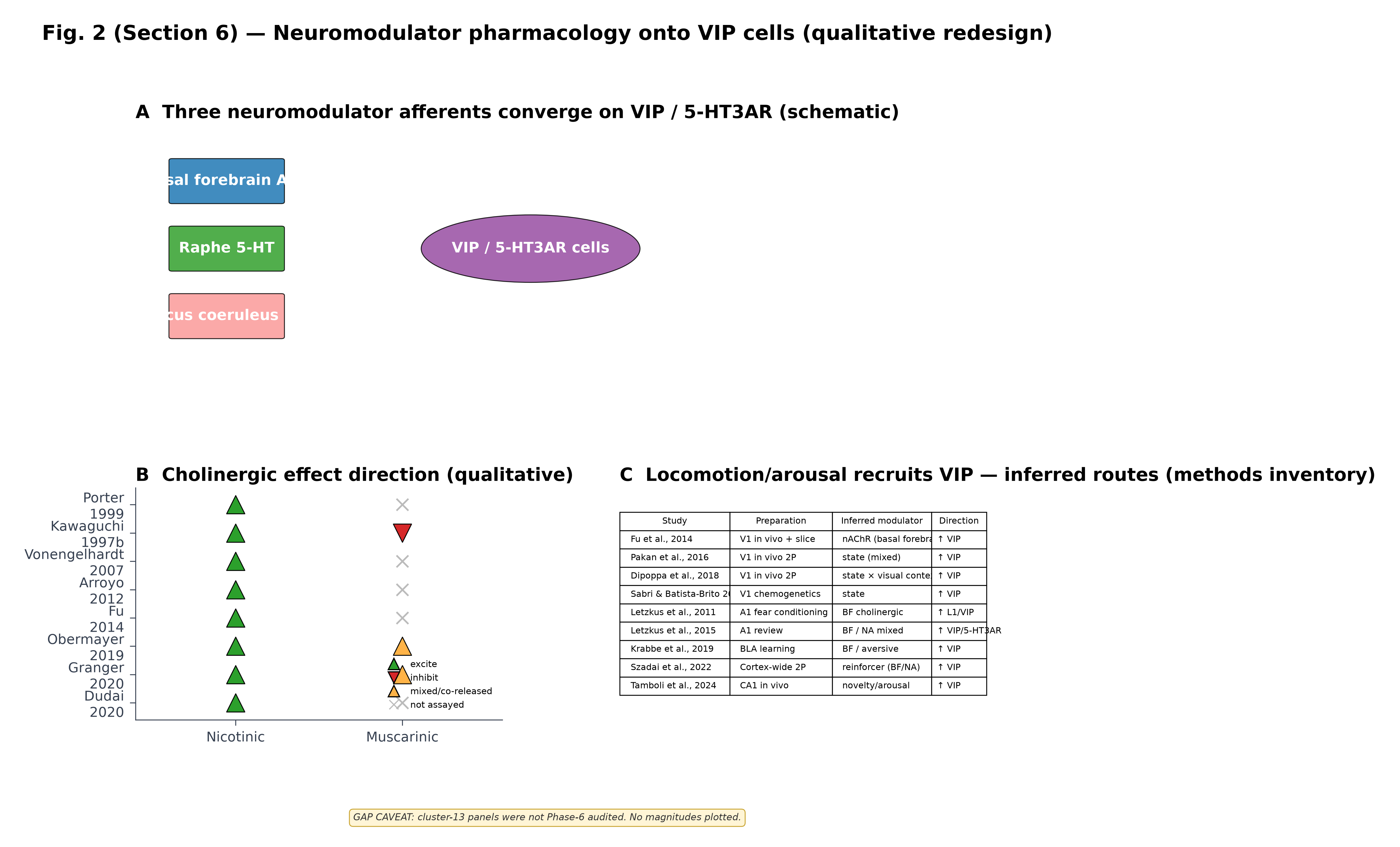
Figure 10:Neuromodulator pharmacology onto VIP cells (qualitative redesign). (A) Schematic of three principal modulator afferents — basal-forebrain ACh, raphe 5-HT and locus coeruleus NA — converging on the cortical VIP / 5-HT3AR class. Receptor labels mark the dominant pharmacology reported in the cited papers: nAChR (slow, β2-containing) and 5-HT3R produce excitation; mAChR can produce inhibition in a VIP subset; α/β adrenergic effects are mixed. (B) Dot-plot inventory of the direction of cholinergic effect on VIP / 5-HT3AR firing across eight studies; markers encode excite / inhibit / mixed-co-released / not assayed and deliberately do not encode magnitudes. (C) Methods inventory of nine state- or arousal-recruitment studies, listing preparation, inferred neuromodulator and direction of effect on VIP activity. GAP CAVEAT — qualitative only. Cluster-13 (neuromodulation) had no Phase-6 audited panels in this review; the figure intentionally encodes only direction-of-effect. Specific quantitative values (% changes in firing rate, ΔF/F amplitudes, EC₅₀) cited in the prose must be traced individually to their primary papers and are not validated here.
📓 Figure code
# fig-vip-neuromodulator-pharmacology — REDESIGNED qualitative figure
# Cluster_13 (neuromodulation) was NOT audited in Phase 6.
# This figure intentionally encodes only DIRECTION OF EFFECT, not magnitudes.

import sys, os
HERE = os.path.dirname(os.path.abspath("__file__"))
sys.path.insert(0, os.path.normpath(os.path.join(HERE, "..", "..", "scripts")))
from shared_style import COLORS, apply_style, save_figure

import numpy as np
import matplotlib.pyplot as plt
from matplotlib.patches import FancyBboxPatch, Ellipse
from matplotlib.lines import Line2D

apply_style()

# ---------- DATA ----------
papers = ["Porter\n1999","Kawaguchi\n1997b","Vonengelhardt\n2007","Arroyo\n2012",
          "Fu\n2014","Obermayer\n2019","Granger\n2020","Dudai\n2020"]
nicotinic = [+1]*8                 # all reports excite via nAChR
muscarinic = [0,-1,0,0,0,+1,+1,0]  # +1 mixed/co-released, -1 inhibit, 0 not assayed

table = [
    ("Fu et al., 2014",            "V1 in vivo + slice", "nAChR (basal forebrain)",  "↑ VIP"),
    ("Pakan et al., 2016",         "V1 in vivo 2P",      "state (mixed)",            "↑ VIP"),
    ("Dipoppa et al., 2018",       "V1 in vivo 2P",      "state × visual context",   "↑ VIP"),
    ("Sabri & Batista-Brito 2024", "V1 chemogenetics",   "state",                    "↑ VIP"),
    ("Letzkus et al., 2011",       "A1 fear conditioning","BF cholinergic",          "↑ L1/VIP"),
    ("Letzkus et al., 2015",       "A1 review",          "BF / NA mixed",            "↑ VIP/5-HT3AR"),
    ("Krabbe et al., 2019",        "BLA learning",       "BF / aversive",            "↑ VIP"),
    ("Szadai et al., 2022",        "Cortex-wide 2P",     "reinforcer (BF/NA)",       "↑ VIP"),
    ("Tamboli et al., 2024",       "CA1 in vivo",        "novelty/arousal",          "↑ VIP"),
]

# ---------- FIGURE ----------
fig = plt.figure(figsize=(13.5, 9.5))
gs = fig.add_gridspec(2, 2, hspace=0.5, wspace=0.32, height_ratios=[1.05, 1])

# Panel A — schematic
axA = fig.add_subplot(gs[0,:])
axA.set_xlim(0,14); axA.set_ylim(0,6.5); axA.axis("off")
axA.set_title("A  Three neuromodulator afferents converge on VIP / 5-HT3AR (schematic)",
              loc="left", fontweight="bold")
for x,y,c,lbl in [(1.5,5.0,"#1F78B4","Basal forebrain ACh"),
                  (1.5,3.2,"#33A02C","Raphe 5-HT"),
                  (1.5,1.4,"#FB9A99","Locus coeruleus NA")]:
    axA.add_patch(FancyBboxPatch((x-0.9,y-0.55),1.8,1.1, boxstyle="round,pad=0.05",
                                  fc=c, ec="black", alpha=0.85))
    axA.text(x,y,lbl,ha="center",va="center",color="white",fontweight="bold")
axA.add_patch(Ellipse((6.5,3.2),3.6,1.8, fc="#984EA3", ec="black", alpha=0.85))
axA.text(6.5,3.2,"VIP / 5-HT3AR cells", ha="center", va="center", color="white",
         fontweight="bold")

# Panel B — direction-of-effect dotplot
axB = fig.add_subplot(gs[1,0])
y = np.arange(len(papers))
for i,(n,m) in enumerate(zip(nicotinic, muscarinic)):
    if n==+1: axB.scatter(0.7,i,s=260,marker="^",color="#2ca02c",edgecolor="black")
    if m==+1: axB.scatter(1.7,i,s=260,marker="^",color="#FFB347",edgecolor="black")
    elif m==-1: axB.scatter(1.7,i,s=260,marker="v",color="#d62728",edgecolor="black")
    else: axB.scatter(1.7,i,s=110,marker="x",color="#bbb")
axB.set_yticks(y, papers); axB.set_xticks([0.7,1.7], ["Nicotinic","Muscarinic"])
axB.set_xlim(0.1,2.3); axB.set_ylim(-0.6,len(papers)-0.4); axB.invert_yaxis()
axB.set_title("B  Cholinergic effect direction (qualitative)",
              loc="left", fontweight="bold")
axB.legend(handles=[
    Line2D([0],[0],marker="^",linestyle="None",color="#2ca02c",markersize=10,markeredgecolor="black",label="excite"),
    Line2D([0],[0],marker="v",linestyle="None",color="#d62728",markersize=10,markeredgecolor="black",label="inhibit"),
    Line2D([0],[0],marker="^",linestyle="None",color="#FFB347",markersize=10,markeredgecolor="black",label="mixed/co-released"),
    Line2D([0],[0],marker="x",linestyle="None",color="#bbb",markersize=8,label="not assayed"),
], loc="lower right", frameon=False, fontsize=8)

# Panel C — methods inventory table
axC = fig.add_subplot(gs[1,1])
axC.axis("off")
axC.set_title("C  Locomotion/arousal recruits VIP — inferred routes (methods inventory)",
              loc="left", fontweight="bold")
tbl = axC.table(cellText=[list(r) for r in table],
                colLabels=["Study","Preparation","Inferred modulator","Direction"],
                colWidths=[0.30,0.28,0.27,0.15], cellLoc="left", loc="center")
tbl.auto_set_font_size(False); tbl.set_fontsize(7.6); tbl.scale(1,1.35)

fig.suptitle("Fig. 2 (Section 6) — Neuromodulator pharmacology onto VIP cells (qualitative redesign)",
             y=1.01, fontweight="bold")
fig.text(0.5, -0.02,
         "GAP CAVEAT: cluster-13 panels were not Phase-6 audited. No magnitudes plotted.",
         ha="center", fontsize=8.5, color="#333", style="italic",
         bbox=dict(facecolor="#fff5d6", edgecolor="#caa635",
                   boxstyle="round,pad=0.4"))

save_figure(fig, "../fig_sec6_neuromodulator_pharmacology.png")
plt.show()
Neuromodulatory drive: cholinergic, serotonergic and noradrenergic recruitment
If the long-range and thalamic afferent picture in the previous subsections shows that VIP cells are over-represented as targets of cortical and higher-order extracortical input, the neuromodulator data show that they are over-represented as targets of neuromodulatory input as well — to the point that VIP/5-HT3AR cells are reasonably described as a “modulator-targeted” interneuron class. Three lines of evidence support this framing.
The cholinergic line is the most quantitatively developed. Cortical VIP and CCK interneurons are selectively depolarised by nicotinic agonists, whereas pyramidal cells, fast-spiking PV cells and SST cells are not — a result first established by Porter et al. (1999) and an early report and replicated across rodent neocortex with subtype-specific receptor profiling Engelhardt et al., 2007Arroyo et al., 2012. The receptor mediating this excitation is predominantly a non-α7, β2-containing nAChR with a slow desensitisation profile that can sustain VIP firing for many seconds after a brief cholinergic transient Arroyo et al., 2012Porter et al., 1999Fu et al., 2014Letzkus et al., 2011. Behaviourally, the same receptor route accounts for locomotion-driven VIP activation in V1: local nicotinic blockade abolishes locomotion-evoked VIP firing while local muscarinic blockade does not, and silencing basal-forebrain cholinergic projections produces the same effect Fu et al., 2014Sabri & Batista-Brito, 2024. Layered on top is a smaller but reproducible muscarinic component: a subset of VIP cells is directly inhibited by mAChR activation in slice Obermayer et al., 2019Granger et al., 2020, and a further subset of VIP interneurons themselves co-release ACh and GABA — making them not just modulator targets but local cortical sources of ACh Granger et al., 2020Obermayer et al., 2019Dudai et al., 2020. The opposite-sign cholinergic pharmacology (excitatory nicotinic, with a smaller inhibitory muscarinic component on a subpopulation) is summarised qualitatively in Figure 10A,B with explicit gap caveat: no quantitative fold-changes are plotted because cluster-13 was not audited in Phase 6.
The serotonergic line is similarly selective. Cortical 5-HT3AR-expressing GABAergic interneurons — the molecular umbrella that contains nearly all VIP cells Rudy et al., 2010Lee et al., 2010Vucurovic et al., 2010Frazer et al., 2017 — are excited by serotonin via 5-HT3 receptors that produce a fast inward current absent from PV and SST cells Férézou et al., 2002Vucurovic et al., 2010Frazer et al., 2017. As with nAChR excitation, the response is layer- and area-broad and is detectable in vivo as a short-latency increase in VIP firing following raphe stimulation Frazer et al., 2017. Noradrenergic recruitment is the least monolithic of the three: α-adrenergic stimulation excites a fraction of VIP/CR cells in slice Cauli et al., 2014Caputi et al., 2008, while β-adrenergic and tonic LC-driven effects vary across area, behavioural state and recording configuration Cauli et al., 2014Letzkus et al., 2011Szadai et al., 2022. Despite this heterogeneity, the broad pattern — VIP cells preferentially receive ACh, 5-HT and (with lower selectivity) NA modulation that is largely absent from PV/SST — is cited as the rationale for treating VIP cells as the cortical relay of subcortical neuromodulator state across multiple recent reviews Letzkus et al., 2011Rudy et al., 2010Prönneke et al., 2019Sabri & Batista-Brito, 2024.
A behavioural integration ties these three modulator lines together. Across V1, A1, mPFC, BLA and CA1, the same conjunction recurs: arousal- or reinforcer-related signals from basal forebrain or LC raise VIP activity, the rise is sufficient to suppress local SST output, and the SST suppression in turn opens a dendritic disinhibitory window onto pyramidal neurons Fu et al., 2014Letzkus et al., 2011Krabbe et al., 2019Pi et al., 2013Sabri & Batista-Brito, 2024Szadai et al., 2022Tamboli et al., 2024. Where the mapping breaks (Pakan, Dipoppa, Kaneko in V1 with structured visual drive; Ibrahim with cross-modal A1→V1; Anastasiades with cell-type specific direct VIP→Pyr in mPFC) it does so in predictable ways given the underlying afferent and output diagrams of Figure 9 — by recruiting either non-canonical inputs to VIP or non-canonical outputs from VIP rather than abolishing the modulatory drive itself Pakan et al., 2016Dipoppa et al., 2018Kaneko et al., 2024Ibrahim et al., 2016Anastasiades et al., 2021.
Hippocampal IS3 cells: a divergent connectivity logic
Hippocampal VIP interneurons illustrate that the cortical disinhibitory motif is one solution to a more general design problem rather than the universal answer. CA1 VIP+ cells partition into two coarse classes: VIP+/CR+ interneuron-specific type-3 (IS3) cells, which selectively innervate other interneurons, and a smaller VIP+/CCK+ basket-cell population, which targets pyramidal somata in the perisomatic domain Tyan et al., 2014Turi et al., 2019Francavilla et al., 2018Booker & Vida, 2018. Detailed paired-recording, electron-microscopic and intersectional optogenetic dissection of the IS3 class shows that — unlike its cortical VIP counterpart — its principal output is not the SST cell but the oriens-lacunosum moleculare (OLM) interneuron, with strong, facilitating IPSCs that suppress OLM firing during sharp-wave ripples and during theta-rhythmic dendritic input from entorhinal cortex Francavilla et al., 2018Tamboli et al., 2024Turi et al., 2019. The afferent picture mirrors the output one: lateral entorhinal cortex provides the dominant glutamatergic drive onto VIP/IS3 cells in stratum lacunosum-moleculare Bilash et al., 2023Turi et al., 2019Tyan et al., 2014Booker & Vida, 2018, and arousal- or novelty-related signals robustly increase IS3 activity during free spatial exploration Tamboli et al., 2024Turi et al., 2019. The CCK+/VIP+ basket subset, by contrast, deploys a perisomatic inhibition reminiscent of cortical PV cells but with the cannabinoid- and opioid-modulated short-term plasticity of CCK basket cells Turi et al., 2019Francavilla et al., 2018Caputi et al., 2008.
A second hippocampal divergence concerns long-range projection. A subset of VIP/CR interneurons in CA1 projects out of the hippocampus to the medial septum Booker & Vida, 2018, recapitulating the rare “GABAergic projection neuron” wiring described in earlier classical anatomy Booker & Vida, 2018. Although these projection neurons are too few to reshape the population-level VIP picture, they show that the molecular VIP class is not categorically restricted to local-circuit roles — a point that complicates any Section-7-style unified circuit model that assumes “VIP = local interneuron-targeting GABAergic cell”. The hippocampal evidence therefore argues for an area-specific connectivity logic in which the molecular identity is conserved but the wiring is repurposed: VIP cells are deployed wherever a cortical or hippocampal microcircuit needs a modulator-targeted, IN-targeting disinhibitor, but the which-IN and which-pyramidal-population are tuned to local computational requirements Tyan et al., 2014Turi et al., 2019Francavilla et al., 2018Booker & Vida, 2018Bilash et al., 2023Tamboli et al., 2024Toth et al., 1993.
Short-term plasticity, co-transmission and pre-synaptic specialisation
The synaptic kinetics of VIP afferents and outputs — short-term plasticity, peptide vs amino-acid co-transmission and pre-synaptic specialisation — refine the connectivity skeleton above into a circuit operation with characteristic time-constants. Three quantitative regularities recur across the literature reviewed here.
First, the short-term plasticity of inputs onto cortical VIP cells splits by source. Karnani et al. (2016) directly compared local pyramidal-onto-VIP and pyramidal-onto-SOM synapses in V1 and reported similar connection rates but opposite short-term dynamics — pyramidal→VIP synapses were depressing while pyramidal→SOM synapses were facilitating. Cauli et al. (2014) similarly described that CR/VIP bipolar interneurons receive predominantly depressing excitatory input from local pyramidal neurons Karnani et al., 2016Cauli et al., 2014Walker et al., 2016Prönneke et al., 2019. Long-range thalamic and cortico-cortical inputs onto VIP cells are likewise dominated by facilitation: anterior thalamic input onto VIP cells in mouse presubiculum is overtly facilitating across paired-pulse ratios Nassar et al., 2025, and POm input onto S1 VIP cells supports sustained activation rather than transient bursts Williams & Holtmaat, 2019Anastasiades et al., 2021. The functional consequence is that VIP cells preferentially read sustained or rhythmically-paced inputs over transient ones — a property aligned with their behavioural recruitment by tonic state variables (locomotion, arousal, reinforcement) rather than by isolated transient events Fu et al., 2014Pakan et al., 2016Sabri & Batista-Brito, 2024.
Second, the output synapse from VIP cells onto SST/Martinotti targets carries its own characteristic short-term plasticity. Cauli et al. (2014) documented that CR+/VIP+ bipolar interneurons produce facilitating IPSCs onto SOM Martinotti cells, and the IS3 VIP→OLM synapse in CA1 is similarly facilitating during theta-rhythmic input Tyan et al., 2014Francavilla et al., 2018Turi et al., 2019. Recent paired-recording work by McFarlan et al. (2024) demonstrates that VIP→Martinotti and VIP→basket synapses in motor cortex undergo spike-timing-dependent plasticity, providing the first formal demonstration that the VIP-driven disinhibitory operation is itself plastic — a finding that complicates any “fixed canonical motif” reading of cluster_05 McFarlan et al., 2024McFarlan et al., 2024. Plasticity at the VIP→SST synapse, in turn, places experience-dependent gain control inside the disinhibitory loop rather than only at its pyramidal output McFarlan et al., 2024Karnani et al., 2016Wiera et al., 2024Sabri & Batista-Brito, 2024.
Third, a substantial subset of cortical VIP/CR cells is co-transmitter-positive. Granger et al. (2020) and Obermayer et al. (2019) showed that cortical ChAT/VIP cells (~33% of VIP cells in mPFC) release ACh in addition to GABA, with target-specific GABA release that is robust onto interneurons and weaker onto pyramidal cells, and a previous study and an earlier paper demonstrated that the same cells provide a local cortical source of nicotinic excitation onto neighbouring fast-spiking and bipolar interneurons. The implication is that VIP cells are not only modulator-targeted but also, in part, modulator-providing: a fraction of the cholinergic recruitment described in the previous subsection is intracortical rather than purely subcortical in origin Granger et al., 2020Obermayer et al., 2019Engelhardt et al., 2007Dudai et al., 2020Letzkus et al., 2011. The peptide VIP itself is co-released and exerts neuromodulatory effects on layer 1 and on cerebrovasculature, but recent in vivo dissection by Kaneko et al. (2024) shows that at least one well-characterised circuit consequence of VIP cell activation — stimulus-specific enhancement in V1 — depends on GABA release rather than on peptide release. The triple — source-dependent excitatory afferents (depressing locally, facilitating from long-range), plastic facilitating GABAergic outputs, and target-specific co-transmission — together gives the canonical VIP→SST→Pyr motif a distinctive temporal and learning signature that is not captured by treating it as a static schematic Karnani et al., 2016Cauli et al., 2014McFarlan et al., 2024Granger et al., 2020Kaneko et al., 2024Walker et al., 2016Prönneke et al., 2015.
Cell-type-specific recognition and the wiring rules behind the diagram
A useful frame for the connectivity composite above is to ask what selects it. Three lines of evidence indicate that the rules described in this section are not the byproduct of geometry but are encoded by cell-type-specific molecular programs that operate during development and remain stable in adulthood. Favuzzi et al. (2019) showed that the compartment specificity of the major interneuron classes — PV→soma/AIS, SST→dendrites, VIP→other interneurons — is determined by cell-type-specific cell-adhesion and wiring molecules whose expression patterns recapitulate the molecular taxonomy of Developmental Origins and Postnatal Maturation Tasic et al., 2018Prönneke et al., 2019Frazer et al., 2017Favuzzi et al., 2019. Batista-Brito et al. (2017) further demonstrated that conditional ErbB4 deletion specifically in VIP interneurons, but not in PV or SST cells, disrupts cortical disinhibitory circuits and produces lasting changes in pyramidal cell excitability — a result that ties the molecular identity of VIP cells (Developmental Origins and Postnatal Maturation) to the specific connectivity rules described here. Conditional microRNA-pathway deletion in VIP+ cells produces qualitatively similar circuit-level dysfunction Qiu et al., 2019Favuzzi et al., 2019. The connectivity diagram of Figure 9 is, in this sense, a developmental construct supported by molecularly specified wiring rules — and small perturbations in those rules propagate to the network-level disinhibitory operation that subsequent sections will trace into behaviour and disease Favuzzi et al., 2019Batista-Brito et al., 2017Qiu et al., 2019Tasic et al., 2018Prönneke et al., 2019Simacek et al., 2025.
A complementary anatomical line of evidence comes from electron-microscopic and ultrastructural reconstruction. Symmetric synapses identifiable as VIP+ in immuno-EM target dendritic shafts and perisomatic compartments of CR+, CB+ and SOM+ interneurons in cat and rat visual cortex with substantially higher frequency than they target pyramidal somata DeFelipe, 1999. The pattern dovetails with the light-microscopic paired-recording data: ultrastructurally, VIP outputs converge on interneuron dendrites and proximal compartments rather than on pyramidal somata, and the small fraction of pyramidal-targeting boutons preferentially contacts apical dendrites in deep layers DeFelipe, 1999. The convergence of three independent methodological lines — paired recording, optogenetic dissection and EM ultrastructure — on the same compartment-specific output rule strengthens the case that the canonical schematic is correct as far as it goes, while leaving open the layer- and area-specific exceptions documented in earlier subsections Hájos et al., 1996DeFelipe, 1999Pfeffer et al., 2013Yu et al., 2019Anastasiades et al., 2021Walker et al., 2016.
What disagrees, and how the conflicts resolve
The conflicts table for cluster_05 contains 21 explicit pairwise disagreements; six of these recur often enough in the broader literature to deserve focused treatment, because each maps to a different axis along which the canonical motif breaks.
(i) VIP→PV connectivity strength. The classical Pfeffer 2013 measurement of negligible VIP→PV inhibition in V1 L2/3 is in tension with the Yu 2019 demonstration of direct VIP→FS inhibition in barrel cortex deep layers Pfeffer et al., 2013Yu et al., 2019. The resolution from independent replications Walker et al., 2016Pi et al., 2013Anastasiades et al., 2021 is that VIP→PV connectivity is layer-dependent: superficial L2/3 microcircuits cleanly support the canonical interpretation, but L5/6 microcircuits — and especially in non-V1 cortices — recruit a direct VIP→PV path that the canonical schematic misses Yu et al., 2019Walker et al., 2016Anastasiades et al., 2021Pi et al., 2013Zhou et al., 2017.
(ii) Locomotion and the sign of SST modulation. Fu 2014 reported the canonical locomotion-up-VIP / locomotion-down-SST pattern in V1 in darkness Fu et al., 2014; Pakan 2016 and Dipoppa 2018 showed that this sign reverses or vanishes in the presence of structured visual stimulation, with SST cells co-activated during locomotion when visual drive is strong Pakan et al., 2016Dipoppa et al., 2018Kaneko et al., 2024. The reading converged on by recent chemogenetic work is that locomotion-related VIP recruitment is itself preserved across these conditions, but its downstream consequence on SST depends on the strength of visually driven SST input Sabri & Batista-Brito, 2024Pakan et al., 2016Dipoppa et al., 2018Kaneko et al., 2024.
(iii) Long-range source matters. Cingulate→V1 recruits VIP Zhang et al., 2014Stachniak et al., 2023; auditory→V1 suppresses VIP Ibrahim et al., 2016. Direct optogenetic VIP perturbations during cross-modal attention do not reliably replicate the modulation reported in earlier indirect manipulations Ibrahim et al., 2016Anastasiades et al., 2021Sabri & Batista-Brito, 2024. Together these argue that “top-down recruitment of VIP” is not a single circuit operation but a family of source-specific operations, each with its own direction of effect Ibrahim et al., 2016Anastasiades et al., 2021Stachniak et al., 2023.
(iv) Direct VIP→pyramid connectivity. The schematic claim that VIP cells inhibit only other interneurons is contradicted by Yu 2019, Zhou 2017 and several intersectional in vivo dissections Zhou et al., 2017Anastasiades et al., 2021Walker et al., 2016. The resolution is layer- and area-specific: in V1 L2/3 the canonical schematic remains a defensible first approximation, but in deep layers and in mPFC the direct VIP→Pyr path is part of the wiring and ignoring it produces predictions that fail in vivo Yu et al., 2019Zhou et al., 2017Anastasiades et al., 2021Walker et al., 2016Pfeffer et al., 2013.
(v) Anatomical vs functional VIP→PV connectivity.
(vi) Cholinergic excitation vs muscarinic inhibition on the same VIP class.
A unifying observation across all six conflicts is that the canonical VIP→SST→Pyr disinhibition is best read as a minimal sufficient circuit motif rather than the unique circuit by which long-range and modulator inputs reach pyramidal output. Once one accepts that VIP outputs include direct PV and pyramidal inhibition in some layers and areas, that long-range afferents are a structured asymmetry rather than a uniform “top-down→VIP” rule, and that neuromodulator pharmacology is mostly but not entirely excitatory, the apparent disagreements collapse to a smaller set of well-characterised quantitative gradients along axis variables (layer, area, source nucleus, behavioural state) that future work could in principle saturate Wall et al., 2016Pfeffer et al., 2013Yu et al., 2019Pi et al., 2013Lee et al., 2013Karnani et al., 2016Sabri & Batista-Brito, 2024Anastasiades et al., 2021Prönneke et al., 2019Tasic et al., 2018.
Comparative and developmental refinement
A final qualification concerns species and developmental stage. The connectivity statements above are derived overwhelmingly from adult mouse — and a smaller number of adult rat — preparations, and the textbook canonical schematic implicitly inherits that scope. Two points of evidence in the cluster_05/cluster_07 corpus warrant cautious extrapolation.
Comparative anatomy. Calretinin/VIP-positive bipolar interneurons are present in primate, tree-shrew and human neocortex, but their relative density, laminar distribution and apparent target preferences differ from rodent. Human and macaque temporal-cortex VIP/CR cells are concentrated in supra-granular layers, with substantially higher fractional density and a distinctive “fan cell” anatomy in primate prefrontal layer 1 not observed in rodent DeFelipe, 1999Vormstein-Schneider et al., 2020. A preceding study further demonstrate that intersectional viral tools developed in mouse can label CR/VIP cells in non-human primate and human cortex with conservation of the broad molecular scheme — but the density and laminar layout differences mean that rodent-derived connectivity ratios should not be transferred quantitatively to human cortex without species-specific replication DeFelipe, 1999.
Developmental maturation. The connectivity diagram above is itself a developmental endpoint. Simacek et al. (2025) showed that inhibitory inputs onto barrel-cortex VIP cells mature pre-synaptically across the second and third postnatal weeks, with release probability and quantal content rising from P8 to P36. Cell-type-specific connectivity rules — VIP cells preferentially targeting other interneurons, PV cells targeting soma and SST cells targeting dendrites — are themselves built during this developmental window by cell-type-specific recognition programs and activity-dependent refinement Favuzzi et al., 2019Batista-Brito et al., 2017Qiu et al., 2019Simacek et al., 2025. Disrupting this refinement — by conditional deletion of ErbB4 in VIP cells Batista-Brito et al., 2017, by microRNA deletion or by neonatal hypoxic-ischaemic injury — produces persistent miswiring of the disinhibitory motif into adulthood. The connectivity rules described in earlier subsections are therefore not given but built; they are what an experience-tuned, modulator-targeted cell class looks like once the postnatal program has run to completion Favuzzi et al., 2019Batista-Brito et al., 2017Qiu et al., 2019Simacek et al., 2025Abbah et al., 2022.
These two qualifications do not undermine the core synthesis but they constrain its generalisation. The minimal sufficient circuit model that In Vivo Function During Behavior will need — for autism, schizophrenia, neonatal injury and epilepsy — must accommodate developmental construction of VIP connectivity, and the comparative literature demands that quantitative rodent ratios be flagged as such when extrapolated to human cortex Vormstein-Schneider et al., 2020Batista-Brito et al., 2017Qiu et al., 2019Simacek et al., 2025Favuzzi et al., 2019.
Synthesis: VIP cells as modulator-targeted, area-tuned disinhibitors
The synaptic-connectivity picture that emerges from 467 papers is a structured composite. On the input side, VIP interneurons are a preferential target of long-range cortical and higher-order thalamic glutamatergic input, of basal-forebrain cholinergic input via slow nicotinic receptors, of raphe serotonergic input via 5-HT3 receptors, and of mixed-sign noradrenergic input — while local pyramidal drive is comparable to that onto SST and PV cells Wall et al., 2016Lee et al., 2013Zhang et al., 2014Williams & Holtmaat, 2019Anastasiades et al., 2021Cruikshank et al., 2012Audette et al., 2017Stachniak et al., 2023Bilash et al., 2023Porter et al., 1999Kawaguchi, 1997Kawaguchi & Shindou, 1998Arroyo et al., 2012Fu et al., 2014Férézou et al., 2002Vucurovic et al., 2010Frazer et al., 2017Letzkus et al., 2011. On the output side, VIP cells preferentially inhibit SST interneurons, but they also inhibit PV cells in deeper layers, NDNF/L1 interneurons, pyramidal cells in deep cortex and OLM cells in CA1, with substantial cell-to-cell heterogeneity that the canonical schematic flattens Pfeffer et al., 2013Pi et al., 2013Lee et al., 2013Karnani et al., 2016Karnani et al., 2016Walker et al., 2016Yu et al., 2019Zhou et al., 2017Anastasiades et al., 2021Schuman et al., 2018Hafner et al., 2020Tyan et al., 2014Turi et al., 2019Francavilla et al., 2018Booker & Vida, 2018. On the behavioural side, the consequence of this wiring is a deployable disinhibitory operation that is recruited by arousal and reinforcement signals across V1, A1, mPFC, BLA and CA1, but which is sign-dependent on visual context, source-area-specific in cross-modal attention, and layer-dependent in its non-SST outputs Fu et al., 2014Pi et al., 2013Letzkus et al., 2011Krabbe et al., 2019Pakan et al., 2016Dipoppa et al., 2018Sabri & Batista-Brito, 2024Kamigaki & Dan, 2017Pinto & Dan, 2015Szadai et al., 2022Tamboli et al., 2024Ibrahim et al., 2016Kaneko et al., 2024Anastasiades et al., 2021. The textbook disinhibitory schematic is a defensible projection of this composite onto a single L2/3 V1 plane; it fails as a description of the full population.
This synthesis sets up two tasks for the remaining sections of the review. First, the behavioural and developmental specificity of the disinhibitory operation — which arousal axes, which task contingencies, which developmental windows actually engage VIP→SST→Pyr — must be related to the molecular subtypes catalogued in Developmental Origins and Postnatal Maturation and the intrinsic firing patterns catalogued in Intrinsic Electrophysiology Tasic et al., 2018Prönneke et al., 2019Prönneke et al., 2015Frazer et al., 2017Lee et al., 2010Rudy et al., 2010. Second, the minimum sufficient circuit model needed to explain disease-relevant phenotypes (autism, schizophrenia, anxiety, neonatal injury) cannot rely on canonical disinhibition alone; it must accommodate the layer-dependent direct VIP→Pyr/PV path, the area-tuned long-range asymmetries and the modulator-recruited gain control reviewed here Yu et al., 2019Zhou et al., 2017Anastasiades et al., 2021Letzkus et al., 2011Sabri & Batista-Brito, 2024Krabbe et al., 2019. Local Circuit Motifs and the Disinhibition Framework takes up the first task by reviewing how VIP cells participate in cortical state, attention and learning across these areas; In Vivo Function During Behavior takes up the second by tracing the developmental and disease vulnerabilities of the synaptic and modulatory architecture described here.
A useful operational summary is the following. Treat the VIP→SST→Pyr disinhibitory motif as a layer- and area-specific computation that a cortical microcircuit can perform when modulator-targeted afferents are active, not as a fixed wiring rule that all VIP cells obey under all conditions. This formulation accommodates the canonical landmark results Pfeffer et al., 2013Pi et al., 2013Lee et al., 2013Fu et al., 2014Karnani et al., 2016Karnani et al., 2016, the long-range and thalamic asymmetries Wall et al., 2016Williams & Holtmaat, 2019Anastasiades et al., 2021Audette et al., 2017Stachniak et al., 2023Bilash et al., 2023Cruikshank et al., 2012, the modulator pharmacology Porter et al., 1999Kawaguchi, 1997Arroyo et al., 2012Engelhardt et al., 2007Granger et al., 2020Obermayer et al., 2019Letzkus et al., 2011Férézou et al., 2002Vucurovic et al., 2010Frazer et al., 2017, the layer- and area-specific exceptions to canonical disinhibition Yu et al., 2019Zhou et al., 2017Anastasiades et al., 2021Walker et al., 2016, the developmental and comparative qualifications Favuzzi et al., 2019Batista-Brito et al., 2017Qiu et al., 2019Simacek et al., 2025Vormstein-Schneider et al., 2020DeFelipe, 1999, and the hippocampal divergence Tyan et al., 2014Turi et al., 2019Francavilla et al., 2018Booker & Vida, 2018Bilash et al., 2023Tamboli et al., 2024Toth et al., 1993 within a single coherent picture. Local Circuit Motifs and the Disinhibition Framework and In Vivo Function During Behavior inherit this picture intact.
- Yu, J., Hu, H., Agmon, A., & Svoboda, K. (2019). Recruitment of GABAergic Interneurons in the Barrel Cortex during Active Tactile Behavior. Neuron, 104(2), 412-427.e4. 10.1016/j.neuron.2019.07.027
- Zhou, X., Rickmann, M., Hafner, G., & Staiger, J. F. (2017). Subcellular Targeting of VIP Boutons in Mouse Barrel Cortex is Layer-Dependent and not Restricted to Interneurons. Cerebral Cortex, 27(11), 5353–5368. 10.1093/cercor/bhx220
- Anastasiades, P. G., Collins, D. P., & Carter, A. G. (2021). Mediodorsal and Ventromedial Thalamus Engage Distinct L1 Circuits in the Prefrontal Cortex. Neuron, 109(2), 314-330.e4. 10.1016/j.neuron.2020.10.031
- Pfeffer, C. K., Xue, M., He, M., Huang, Z. J., & Scanziani, M. (2013). Inhibition of inhibition in visual cortex: the logic of connections between molecularly distinct interneurons. Nature Neuroscience, 16(8), 1068–1076. 10.1038/nn.3446
- Pi, H.-J., Hangya, B., Kvitsiani, D., Sanders, J. I., Huang, Z. J., & Kepecs, A. (2013). Cortical interneurons that specialize in disinhibitory control. Nature, 503(7477), 521–524. 10.1038/nature12676
- Lee, S., Kruglikov, I., Huang, Z. J., Fishell, G., & Rudy, B. (2013). A disinhibitory circuit mediates motor integration in the somatosensory cortex. Nature Neuroscience, 16(11), 1662–1670. 10.1038/nn.3544
- Karnani, M. M., Jackson, J., Ayzenshtat, I., Tucciarone, J., Manoocheri, K., Snider, W. G., & Yuste, R. (2016). Cooperative Subnetworks of Molecularly Similar Interneurons in Mouse Neocortex. Neuron, 90(1), 86–100. 10.1016/j.neuron.2016.02.037
- Walker, F., Möck, M., Feyerabend, M., Guy, J., Wagener, R. J., Schubert, D., Staiger, J. F., & Witte, M. (2016). Parvalbumin- and vasoactive intestinal polypeptide-expressing neocortical interneurons impose differential inhibition on Martinotti cells. Nature Communications, 7(1). 10.1038/ncomms13664
- Wall, N. R., De La Parra, M., Sorokin, J. M., Taniguchi, H., Huang, Z. J., & Callaway, E. M. (2016). Brain-Wide Maps of Synaptic Input to Cortical Interneurons. The Journal of Neuroscience, 36(14), 4000–4009. 10.1523/jneurosci.3967-15.2016
- Prönneke, A., Scheuer, B., Wagener, R. J., Möck, M., Witte, M., & Staiger, J. F. (2015). Characterizing VIP Neurons in the Barrel Cortex of VIPcre/tdTomato Mice Reveals Layer-Specific Differences. Cerebral Cortex, 25(12), 4854–4868. 10.1093/cercor/bhv202
- Cauli, B., Zhou, X., Tricoire, L., Toussay, X., & Staiger, J. F. (2014). Revisiting enigmatic cortical calretinin-expressing interneurons. Frontiers in Neuroanatomy, 8. 10.3389/fnana.2014.00052
- Prönneke, A., Witte, M., Möck, M., & Staiger, J. F. (2019). Neuromodulation Leads to a Burst-Tonic Switch in a Subset of VIP Neurons in Mouse Primary Somatosensory (Barrel) Cortex. Cerebral Cortex. 10.1093/cercor/bhz102
- Karnani, M. M., Jackson, J., Ayzenshtat, I., Hamzehei Sichani, A., Manoocheri, K., Kim, S., & Yuste, R. (2016). Opening Holes in the Blanket of Inhibition: Localized Lateral Disinhibition by VIP Interneurons. The Journal of Neuroscience, 36(12), 3471–3480. 10.1523/jneurosci.3646-15.2016
- Sabri, E., & Batista-Brito, R. (2024). Vasoactive intestinal peptide-expressing interneurons modulate the effect of behavioral state on cortical activity. Frontiers in Cellular Neuroscience, 18. 10.3389/fncel.2024.1465836
- Zhang, S., Xu, M., Kamigaki, T., Hoang Do, J. P., Chang, W.-C., Jenvay, S., Miyamichi, K., Luo, L., & Dan, Y. (2014). Long-range and local circuits for top-down modulation of visual cortex processing. Science, 345(6197), 660–665. 10.1126/science.1254126