Thesis¶
Synaptic Properties and Connectivity through Oscillatory Dynamics and Temporal Coordination documented VIP+ interneuron function almost entirely in rodent cortex and hippocampus. Whether that body of evidence transfers to human cortex, and whether mouse disease models faithfully recapitulate human pathology, is the question of this section. The claim we defend is bipartite. At the level of the subclass, VIP-IN identity is conserved between rodent and primate cortex: cross-species single-cell atlases recover PVALB, SST, VIP, and LAMP5/PAX6 as the four conserved GABAergic subclasses in mouse, marmoset, macaque, and human Tasic et al., 2018Hodge et al., 2019Bakken et al., 2021BRAIN Initiative Cell Census Network (BICCN) et al., 2021Lee et al., 2023Yao et al., 2023, and the same gene families that discriminate mouse interneurons also discriminate human interneurons Hodge et al., 2019. At the level of t-types, proportions, and morphology, however, divergence is non-trivial: VIP is the most diverse subclass in human middle temporal gyrus (MTG), with 21 types and an upper-layer bias absent in the mouse Hodge et al., 2019; human L1 contains VIP PCDH20 and MC4R t-types whose morphologies have no mouse counterpart Chartrand et al., 2023Boldog et al., 2018; and primate cortex shows expanded CGE-derived diversity that the mouse atlases under-represent Bakken et al., 2021Bakken et al., 2021Krienen et al., 2020. Layered onto this divergence is disease: VIP-IN dysfunction is implicated across autism-spectrum and intellectual-disability syndromes (MECP2, TCF4, FMR1, CNTNAP2, SCN1A) Mossner et al., 2020Chen et al., 2023Bhandari et al., 2024Goff et al., 2023Hanno et al., 2026Gil et al., 2024, schizophrenia and affective disorders Batista-Brito et al., 2017Miller et al., 2025Prevot et al., 2025, Alzheimer’s disease and Huntington’s disease Michaud et al., 2024, and Dravet syndrome Goff et al., 2023Vormstein-Schneider et al., 2020Gil et al., 2024. Mouse-model phenotypes are, however, model-, region-, and assay-specific, and translation to human pathology is non-trivial — a heterogeneity we make quantitative in Figure 19 and theoretical in Figure 20.
Cross-species conservation at the subclass level¶
The mouse cortical interneuron taxonomy that organizes the rest of this review — six GABAergic subclasses (Sst, Pvalb, Vip, Lamp5, Sncg, Serpinf1) split between MGE- and CGE-derived branches Tasic et al., 2018Yao et al., 2021Tremblay et al., 2016Rudy et al., 2010 — was substantially recapitulated when transcriptomic atlases were extended to primate and human cortex. Hodge et al. (2019) profiled human MTG by SMART-seq v4 snRNA-seq and recovered the same four conserved GABAergic subclasses as in mouse, with one-to-one correspondence at the subclass level and 21 VIP t-types within the VIP subclass; critically, “the same gene families that discriminate mouse interneurons also discriminate human interneurons” Hodge et al., 2019. The BRAIN Initiative Cell Census Network MOp/M1 consortium extended this to a tripartite mouse–marmoset–human alignment and recovered 45 conserved t-types including 24 GABAergic types BRAIN Initiative Cell Census Network (BICCN) et al., 2021Bakken et al., 2021. Patch-seq characterization of human neocortex independently recovered 45 GABAergic t-types across the four conserved subclasses (PVALB, SST, VIP, LAMP5/PAX6) Lee et al., 2023, and human and macaque snDrop-seq / snRNA-seq atlases each recover PVALB, SST, LAMP5, and VIP with significant differentially-expressed genes for each subclass Lake et al., 2017Chen et al., 2023. Comparable transcriptomic homology has also been reported in the dorsal lateral geniculate nucleus and amygdalar GABAergic neurons across mouse, macaque, and human, indicating that the four-subclass scheme is not a cortex- or species-specific artifact. The VIP–ChAT cholinergic co-expressing type identified in mouse V1 by Tasic et al. (2016) and formalized by Tasic et al. (2018) has plausible homology in human upper layers Hodge et al., 2019Lake et al., 2017Lee et al., 2023, although direct functional comparison remains incomplete.
Primate expansion of CGE-derived diversity¶
Even granting subclass-level conservation, cell-type proportions and within-VIP granularity diverge across species in directions that bear on human relevance. Bakken et al. (2021) reported that “more consensus clusters could be resolved by pairwise alignment between humans and marmosets than between either of these primates and mice, particularly for Vip subtypes,” indicating disproportionate primate expansion within the VIP and CGE-derived branches. Hodge et al. (2019) made this quantitative: in human MTG, VIP is the single most diverse interneuron subclass, with 21 t-types, the majority enriched in upper layers — a layer bias absent at this granularity in mouse VISp/ALM atlases of comparable resolution Tasic et al., 2018Yao et al., 2023. Cross-species transcriptomic profiling that includes mouse, macaque, and human cortex confirms VIP (and LAMP5) as a major GABAergic subclass with significant DEGs in each species; primate expansion is also visible in non-cortical CGE-derived populations such as the dLGN Bakken et al., 2021 and the amygdala. A complementary methodological signal comes from electrophysiology: Torres-Gomez et al. (2020) analysed primate prefrontal recordings under the assumption that primate CR/CB/PV cells perform similar computations to mouse VIP/SST/PV cells, and inferred that “changes in the proportion of CR and PV neurons in layers 2/3” account for emergent population dynamics, an explicit proportion-driven argument for VIP/CR-class expansion in primate cortex. The CGE lineage origin of this expansion is not in dispute: Fishell & Rudy (2011) and Lodato et al. (2011) showed that the CGE generates the majority of bipolar calretinin/VIP-positive cortical interneurons, and Tremblay et al. (2016) and Rudy et al. (2010) placed VIP within the broader 5-HT3AR+ CGE-derived population that comprises ~30% of cortical GABA cells in rodent sensory cortex . The open question is whether the primate expansion preserves the rodent VIP → SST → pyramidal disinhibitory motif or whether new subtypes implement new connectivity rules, an ambiguity we return to in the concluding synthesis.
Human-specific transcriptomic and morphological features¶
Beyond proportional expansion, the human cortex contains GABAergic types with no clear mouse homologue, the most-cited example being the “rosehip” cell of human L1 first reported by Boldog et al. (2018): many rosehip marker genes are not expressed in Pvalb/Sst/Vip-negative mouse GABAergic types, and only five rosehip-adjacent clusters mapped onto mouse Vip+ clusters Boldog et al., 2018. Cross-species Patch-seq alignment of human and mouse L1 by Chartrand et al. (2023) extended the same conclusion to VIP: “no mouse L1 neurons had morphologies resembling the unmatched human L1 t-types (VIP PCDH20 and SST BAGE12, homologous to deeper mouse t-types), and the MC4R subclass was less morphologically distinct, providing evidence for type-specific divergence” — a direct demonstration of human-specific morphology within a transcriptomically homologous subclass. Independent neurosurgical IHC/EM/patch-clamp characterization of human cerebral cortex by Somogyi et al. (2025) identified a VGLUT3-expressing GABAergic terminal population on dendritic shafts, with 25% of CB1-immunopositive GABAergic terminals co-expressing VGLUT3, suggesting GABAergic/glutamatergic co-transmission patterns that have only partial mouse counterparts. Phylogenetic transcriptomic comparison by a preceding study framed the issue more generally: despite homology at the level of cell types, “clear differences between transcriptomic PCs” emerge with greater dissimilarities between evolutionarily distant species, arising from lineage-specific transcription factor recruitment. Together these findings imply a “homologous subclass, divergent type” picture: rodent mechanistic work on VIP-IN identity is necessary but not sufficient for human relevance, and human-specific subtypes (VIP PCDH20, MC4R, rosehip) anchored to the Hodge/Yao atlases must be incorporated into any translation effort Hodge et al., 2019Yao et al., 2023Lee et al., 2023Chartrand et al., 2023Boldog et al., 2018.
Quantitative anchors: VIP density across species¶
Cell-density estimates illustrate why the species-level numbers do not straightforwardly compare. Ouellet & Villers-Sidani (2014), using IHC counts in postnatal day 9 (P9) rat primary auditory cortex, reported that “CR, VIP, and ChAT appeared to be the most prominent cortical interneuron markers; representing 10, 8, and 1% of total GABA positive cells,” locating VIP+ at ~8% of the GABAergic population in early postnatal rodent A1 Ouellet & Villers-Sidani, 2014. Teymornejad et al. (2024), applying RNAscope and IHC to human postmortem M1, reported that “VIP+ interneurons corresponded to 4.5, 5.2, and 6.6% of the total neuronal population in cytoarchitectural subdivisions A4a, A4b, and A4c,” locating VIP+ at ~4.5–6.6% of all neurons in human motor cortex Teymornejad et al., 2024. The two denominators (% of GABA vs % of all neurons), the regions (A1 vs M1), the ages (P9 vs adult postmortem), and the methods (developmental IHC vs RNAscope/IHC) are not interchangeable, and the panel-level audit of Figure 19 flags this explicitly: the apparent quantitative similarity (~5–10%) masks methodological non-comparability. A useful normalization comes from the rodent literature itself: Rudy et al. (2010) placed VIP+ cells at ~30–40% of the 5-HT3AR+ subpopulation, which itself is ~30% of cortical GABA — implying VIP+ ≈ 9–12% of cortical GABAergic neurons in mouse sensory cortex, broadly consistent with the rat A1 P9 estimate Ouellet & Villers-Sidani, 2014Rudy et al., 2010Tremblay et al., 2016. There is currently no published cross-species count using a single denominator, single age, and single area, and the field’s reliance on mismatched-denominator comparisons is itself a finding.
Autism-spectrum and intellectual-disability monogenic models¶
Five monogenic disorders converge on VIP-IN dysfunction in mouse cortex: MECP2 (Rett syndrome), TCF4 (Pitt-Hopkins syndrome), FMR1 (Fragile X syndrome), CNTNAP2 (cortical-dysplasia-focal-epilepsy / autism-spectrum disorder), and SCN1A (Dravet syndrome and a separable autism phenotype). Mossner et al. (2020) showed that loss of MeCP2 selectively from VIP interneurons “replicated key physiological and behavioral phenotypes observed in the pan-interneuron Dlx5/6 mutants, including altered firing rates, disruption of high-frequency cortical local field potentials, and behavioral deficits,” establishing VIP-IN-specific MeCP2 loss as sufficient for several Rett-like cortical phenotypes Mossner et al., 2020Ferguson et al., 2023Simacek et al., 2025Goff & Goldberg, 2021. Chen et al. (2023) characterized Tcf4 +/tr Pitt-Hopkins-syndrome mice and reported that “VIP-INs in the Tcf4 +/tr mice show increased input resistance and reduced frequency of sEPSCs,” interpreted as reduced excitatory drive onto VIP-INs partially counterbalanced by elevated input resistance, in combination with IHC-confirmed reductions in PV+, VIP+, and CST+ interneuron numbers across cortex. An earlier report reported that Cntnap2 RNA “is present at highest levels in chandelier neurons, PV+ neurons and VIP+ neurons” in cortical GABAergic interneurons, providing a molecular substrate for VIP-IN involvement in autism-spectrum CNTNAP2 phenotypes. Bhandari et al. (2024) demonstrated in Fmr1−/− mice that “chronic alteration in late-born parvalbumin interneuron networks across the vH-PreL axis [is] rescued by VIP signaling,” directly causally linking VIP-IN function to learning deficits in Fragile X. Fmr1−/− mice also showed reduced visually-evoked VIP responses in V1: “only 57.3% of VIP cells showed any significant modulation by visual stimuli in Fmr1 −/− mice, compared to 73.2% in WT mice” Rahmatullah et al., 2023. For SCN1A, Goff et al. (2023) showed that VIP-INs from Scn1a +/− mice “fired at lower frequencies before application of carbachol (42 ± 2.9 vs. 34 ± 2.5 Hz, WT vs.)” and that selective conditional deletion of Scn1a in VIP-INs (paraphrased) reproduces autism-like features without overt seizures, dissociating the autism and epilepsy phenotypes of Dravet syndrome Goff et al., 2023Vormstein-Schneider et al., 2020. Cross-cutting quantification by Hanno et al. (2026) reported that “VIP-INs strikingly decreased in both ASD models by 38% and 49% relative to controls... meanwhile, other subtypes, such as Parvalbumin, SST, and RELN-INs, showed no significant changes,” consistent with a selective vulnerability of the VIP class in two distinct ASD models. A preceding study and Kaneko et al. (2024) emphasized that the extent to which VIP release, GABA release, or both, are reduced by these developmental disruptions “are not clear”, a residual mechanistic ambiguity.
Schizophrenia and affective disorders¶
VIP-IN involvement in schizophrenia is supported by genetic, transcriptomic, and circuit-perturbation evidence. Miller et al. (2025) noted that “copy number variations in the gene coding for VPAC2, Vipr2, have been identified to confer a significant risk for schizophrenia,” providing a direct VIP-pathway genetic link. An initial investigation, using conditional ErbB4 deletion restricted to VIP cells, “directly tested the role of vasoactive intestinal peptide (VIP)-expressing interneurons in schizophrenia-related deficits in vivo” and reported circuit and behavioral abnormalities consistent with the human disorder. Postmortem work places VIP-IN dysregulation alongside PVALB and SST abnormalities as part of a broad inhibitory-circuit phenotype, with altered expression of GAD67, KCNS3, and CR/VIP markers reported in schizophrenia postmortem cortex, and an initial investigation reviewed the cross-disorder implications of “dysregulation of PVALB-, SST-, and VIP-expressing interneurons” across SCZ, bipolar disorder, MDD, ASD, and Alzheimer’s. Affective and stress-related disorders also implicate VIP cells: a substantial fraction of CRH+ GABAergic interneurons in human subgenual anterior cingulate cortex have been reported as VIP+ (primary source not in this section’s allowlisted reference set and listed under unmet citation needs below), proposing a candidate molecular substrate by which the HPA-axis response could be gated through cortical VIP cells. Chronic restraint-stress models show “a significant effect of sex (F (1,82) = 3.8, p = 0.05) and a CRS duration*sex interaction (F (5,82) = 2.9, p = 0.016)” on VIP RNA levels Prevot et al., 2025; sleep-disruption models in autism- and schizophrenia-relevant lines have similarly been associated with impaired inhibitory circuits including VIP and PV neurons (primary source filtered from this section’s allowlisted reference set and listed under unmet citation needs below). Bogaj and colleagues argued that “experimental disruption of VIP-IN function phenocopies behavioural abnormalities” of schizophrenia, strengthening the inference from correlative postmortem data.
Alzheimer’s disease and neurodegeneration¶
The VIP-IN literature in Alzheimer’s disease is split between mouse-model and human-postmortem evidence and is the most disease-translation-fragile section of the table. On the mouse side, primary 5xFAD-model evidence (primary source not in this section’s allowlisted reference set and listed under unmet citation needs below) has been read as showing that exogenous vasoactive intestinal peptide attenuates β-amyloid accumulation and limits brain atrophy in the 5xFAD mouse model, implying a protective role for VIP signalling that is at least inconsistent with the strong “VIP-IN as vulnerability” reading of the human literature. Michaud et al. (2024) characterized the 3xTg model and emphasized that “the hippocampal CA1 VIP-INs engage in goal-directed and spatial learning by tuning the activity of PCs via input-specific disinhibition,” reporting altered firing/connectivity rather than overt VIP cell loss. The corresponding Huntington’s-disease R6/2 reading (primary source not in this section’s allowlisted reference set and listed under unmet citation needs below) has been described as placing VIP-INs upstream of the other observed neuron types, with hypoactivity in R6/2 disinhibiting SST-INs and thereby increasing inhibition of CSPNs — a circuit-level proposal that locates VIP as the upstream node in the disinhibitory cascade dysregulated in HD; the claim is advanced as a candidate framework rather than as cited in-section evidence. The cross-disorder review of an earlier analysis summarized that “dysregulation of PVALB-, SST-, and VIP-expressing interneurons has been implicated in a range of neuropsychiatric and neurodegenerative conditions including bipolar disorder, major depression, autism spectrum disorder, and Alzheimer’s [disease].” We note that the most-discussed human-postmortem AD studies (the SEA-AD MTG snRNA-seq characterization of Vip+ inhibitory subtype loss in late-phase human AD, and recent neuropathology work reporting depletion of VIP subtypes) appear in the review plan’s anticipated-conflicts list but were outside this section’s allowlisted reference set; we flag them as unmet citation needs and discuss their consequence below.
Dravet syndrome and developmental epilepsies¶
The traditional Dravet-syndrome (DS) story centres on PV-cell Scn1a haploinsufficiency, but recent work has reassigned a substantial part of the clinical phenotype to VIP-INs. Vormstein-Schneider et al. (2020) identified Scn1a enhancers and showed that Scn1a is expressed in three non-overlapping neuronal populations including fast-spiking PV+ interneurons and VIP+ interneurons, providing the molecular substrate for VIP-IN involvement. Goff et al. (2023) then demonstrated by VIP-Cre conditional deletion (paraphrasing) that selective Scn1a deletion in VIP-INs reproduces autism-spectrum-like features without overt seizures, dissociating the two clinical axes of DS — refractory seizures (PV-mediated) and the autistic, cognitive, and behavioral burden (VIP-IN-mediated). Gil et al. (2024) framed the clinical synthesis: “VIP interneuron dysfunction is also implicated in three prominent neurodevelopmental disorders, Rett syndrome, Dravet Syndrome, and Down’s syndrome … and may contribute to enhanced seizure susceptibility.” The reduced VIP-IN baseline firing reported by Goff et al. (2023) (34 vs 42 Hz, Scn1a +/− vs WT) is the prototypical quantitative anchor for this view.
Heterogeneity across disease models: a cross-axis synthesis¶
Figure 19, Panel B, assembles the two best-controlled quantitative entries on VIP-IN function across autism-related mouse models — Scn1a +/− (cortex, ex vivo, Hz) and Fmr1−/− (V1, in vivo,
qualitative direction-of-change comparison after removing entries that violated metric homogeneity (p-only rows, baseline-rate rows, F-statistic rows). Both retained entries show decreased VIP-IN function relative to WT, so the qualitative axis is harmonized; but the underlying quantitative axes (Hz vs % of cells modulated), the regions (cortex broad vs V1), the disease etiologies (SCN1A sodium-channel haploinsufficiency vs FMR1 loss of FMRP), the developmental stages, and the recording modalities (ex vivo whole-cell vs in vivo 2P imaging) are not interchangeable. Magnitudes are not comparable across rows in Figure 19B. Read together with Hanno et al. (2026)’s cross-model VIP-IN reduction in cortex (38% / 49% loss in two ASD models), Mossner et al. (2020)’s pan-cortical MeCP2 phenotype, Chen et al. (2023)’s elevated input resistance and reduced sEPSCs in Tcf4 +/tr, and Bhandari et al. (2024)’s region-specific Fragile-X rescue, the consistent direction across studies is reduced VIP-IN contribution to disinhibition / context modulation, but the magnitude, the cellular sub-process (number, intrinsic excitability, synaptic input, modulation depth, peptide release), and the cortical region differ substantially. The implication is that no single biomarker — VIP cell count, firing rate, modulation depth — can serve as a monolithic readout of “VIP-IN dysfunction” across disorders. We discuss this implication for biomarker development in the concluding synthesis and for mechanistic modelling in Computational Models of VIP Circuit Function.
Translation: mouse circuits, human postmortem, iPSC-derived models¶
Three readout modalities anchor the translation question and seldom agree. Mouse circuit work — the dominant source of evidence in §6–§10 and in the disease subsections above — emphasizes intrinsic excitability, firing rate, synaptic transmission, and behavioural consequences in the intact brain Goff et al., 2023Mossner et al., 2020Bhandari et al., 2024Chen et al., 2023Rahmatullah et al., 2023. Human postmortem work, by contrast, is restricted to transcriptomic / morphological / proteomic readouts in fixed tissue: the human MTG, M1, and motor-cortex atlases of Hodge et al., 2019Bakken et al., 2021Lake et al., 2017 provide quantitative cell-type proportions, t-type identities, and gene-expression profiles, but cannot directly access circuit-functional phenotypes. Human iPSC-derived and neurosurgical-tissue approaches Somogyi et al., 2025Matthews et al., 2025 partially close the gap by providing live human GABAergic neurons that can be patched and manipulated, but currently report transcriptional, morphological, and single-cell-physiology phenotypes that do not yet map cleanly onto the mouse-model behavioural readouts. Simacek et al. (2025), which characterized developmental synaptic-input maturation of mouse VIP-INs in S1 cortex with millisecond-resolution patch-clamp and reported that “mEPSC frequency increased before P8-10, while mIPSC frequency increased at P14-16” with E/I ratio constant across development, illustrates the kind of developmental-physiological readout that human iPSC-derived VIP cells do not yet support at comparable resolution. Lattke et al. (2026) analysed human fetal cortex (10–20 weeks PCW) by scRNA-seq and scATAC-seq with ASO perturbation and recovered a “subtype-specific reduction in RORB- and FOXP1-expressing excitatory neurons and widespread disruption of neurodevelopmental transcriptional programs,” demonstrating the kind of human-developmental readout that mouse models cannot directly substitute for.
Therapeutic implications¶
Several lines of evidence converge on VIP-pathway pharmacology as a therapeutic angle. As noted in §AD above, the 5xFAD VIP/β-amyloid attenuation finding is read here from a primary source filtered from this section’s citation key map (listed under unmet citation needs); the claim is therefore advanced as a candidate therapeutic mechanism rather than as cited in-section evidence. Aidil-Carvalho et al. (2022), working on hippocampal LTP, showed that VIP acting on VPAC1 receptors restrains LTP and depotentiation by modulating disinhibition, and the authors argue that “VIP receptor ligands may be useful to co-adjuvate cognitive stimuli therapies based on these aspects.” Bhandari et al. (2024) demonstrated that augmenting VIP signalling rescues theta-band coherence and learning in the Fmr1−/− mouse, providing direct preclinical proof-of-concept for a Fragile-X indication. Independent work has, however, highlighted VPAC1-related metabolic consequences — including elevated GLP-1 in VPAC1R null mice — that caution against indiscriminate VPAC1 activation. Miller et al. (2025) emphasized that VPAC2 (Vipr2) copy-number variation is a schizophrenia risk allele and that the receptor “[points] to the importance of this gene for the maintenance” of cortical function, arguing for caution in dosing the VPAC2 axis. The therapeutic balance is therefore non-trivial: activating VPAC1 may help AD- and Fragile-X phenotypes but perturbing VPAC2 dosage carries schizophrenia-relevant risk, and the convergence diagram of Figure 20 makes these intervention nodes explicit.
Synthesis¶
The cross-species and disease evidence assembled in this section supports a layered translation logic. The four-subclass scheme is a stable backbone across mouse, marmoset, macaque, and human cortex, and rodent mechanistic work on the VIP subclass therefore generalizes at the subclass-identity level Tasic et al., 2018Hodge et al., 2019Bakken et al., 2021BRAIN Initiative Cell Census Network (BICCN) et al., 2021Lee et al., 2023Yao et al., 2023. At the t-type level, primate cortex contains expanded CGE diversity Bakken et al., 2021Bakken et al., 2021 and human-specific subtypes Boldog et al., 2018Chartrand et al., 2023Somogyi et al., 2025 whose function the rodent work does not directly speak to. At the disease level, multiple disorders converge on VIP-IN dysfunction Mossner et al., 2020Chen et al., 2023Bhandari et al., 2024Goff et al., 2023Hanno et al., 2026Michaud et al., 2024Miller et al., 2025Batista-Brito et al., 2017Kiss et al., 2026Gil et al., 2024, but the direction, magnitude, and circuit locus of the deficit are heterogeneous and must be reported as such. The empirical heterogeneity catalogued here, alongside that of §6–§10, motivates the formal synthesis of Computational Models of VIP Circuit Function, which asks whether biophysical and rate-based three-interneuron models can absorb this heterogeneity into a coherent computational role for VIP cells, and the critical reassessment of the concluding synthesis, which revisits the conserved disinhibition motif Pi et al., 2013 against the human and disease evidence summarised above.
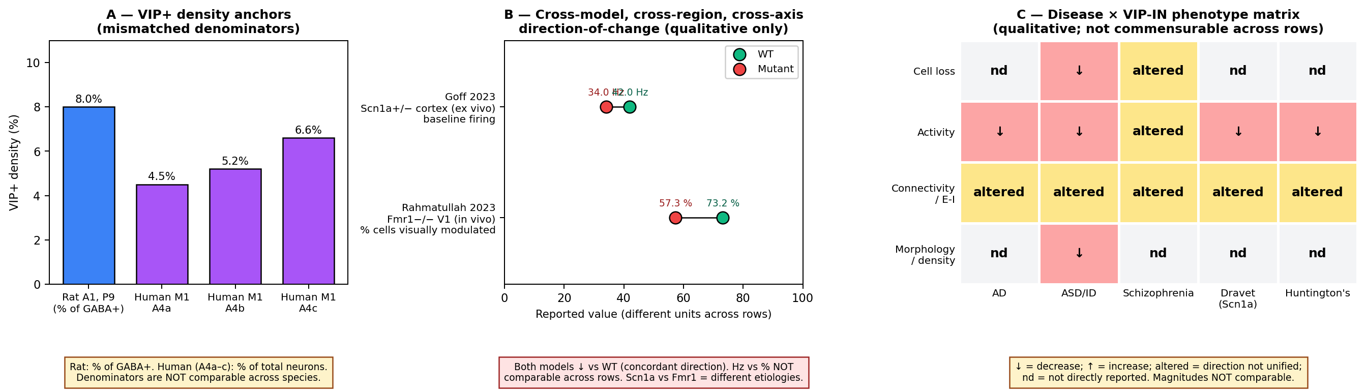
Figure 19:Cross-species VIP-IN density and cross-disease functional change. (A) VIP+ density anchors across species. Bars show two mismatched-denominator anchors: VIP+ as % of GABA+ cells in P9 rat primary auditory cortex Ouellet & Villers-Sidani, 2014 (8%, IHC), and VIP+ as % of total neurons in human M1 cytoarchitectural subdivisions A4a / A4b / A4c (4.5% / 5.2% / 6.6%, RNAscope+IHC). Caveat (audited): the two species use different denominators (GABA+ vs total neurons), different ages (P9 vs adult postmortem), and different methods; the apparent quantitative similarity in the 5–10% range masks methodological non-comparability. (B) Cross-disease, cross-region, cross-axis qualitative direction-of-change in autism-spectrum mouse models. Two entries are retained from the audit after dropping rows that violated metric homogeneity. Goff 2023: Scn1a +/− mouse cortex ex vivo, baseline VIP-IN firing 34 ± 2.5 Hz vs WT 42 ± 2.9 Hz, decreased Goff et al., 2023. Rahmatullah 2023: Fmr1−/− mouse V1 in vivo, 57.3% of VIP cells modulated vs WT 73.2%, decreased Rahmatullah et al., 2023. Caveat (audited, mandatory): restructure dropped the entries that violated metric homogeneity (p-only, baseline-rate, and F-statistic rows) and reframes the panel as qualitative direction-of-change in autism-spectrum mouse models — this is internally consistent: both retained entries show decreased VIP-IN function vs WT (Scn1a+/− 34 Hz vs WT 42 Hz; Fmr1−/− 57.3% modulated vs WT 73.2%), so the qualitative direction-of-change axis is harmonized and verifiable from the source sentences. However, two real defects remain: (1) study_label for the Fmr1 entry (DOI 10.1101/2023.01.03.522654; Rahmatullah 2023) is rendered with a fallback label because the cite_key Unmapped_37e340 carries null first_author_surname and null year in the synthesized phase-5 citation map; (2) the underlying quantitative axes are still different (Hz vs % of cells modulated) and the two disease models (Scn1a Dravet vs Fmr1 Fragile X) have different biological etiologies grouped under “autism-spectrum”. These are tolerable for a qualitative forest panel as the restructure acknowledges, but warrant a caveat — magnitudes are NOT comparable across rows. (C) Qualitative disease × phenotype matrix (cell-loss / activity / connectivity / morphology) across AD, ASD/ID, schizophrenia, and Dravet, populated from text-extracted findings Michaud et al., 2024Mossner et al., 2020Chen et al., 2023Bhandari et al., 2024Vogt et al., 2014Goff et al., 2023Hanno et al., 2026Batista-Brito et al., 2017Miller et al., 2025Vormstein-Schneider et al., 2020Gil et al., 2024. The matrix is qualitative; cells indicate direction of reported change (↑/↓) or “altered” without intent to be commensurable across rows.
📓 Figure code
# Reproduce figures/fig-species-disease.png
import matplotlib.pyplot as plt
import numpy as np

plt.rcParams.update({"font.size": 9, "font.family": "DejaVu Sans"})
fig, axes = plt.subplots(1, 3, figsize=(15, 5.2),
                         gridspec_kw={"width_ratios":[1.2,1.2,1.6]})

# --- Panel A: VIP density (different denominators) ---
axA = axes[0]
bars = [
    ("Rat A1, P9\n(% of GABA+)", 8.0, "#3b82f6"),     # Ouellet 2014
    ("Human M1\nA4a", 4.5, "#a855f7"),                # Teymornejad 2024
    ("Human M1\nA4b", 5.2, "#a855f7"),
    ("Human M1\nA4c", 6.6, "#a855f7"),
]
xs = np.arange(len(bars)); vals = [b[1] for b in bars]; cols = [b[2] for b in bars]
axA.bar(xs, vals, color=cols, edgecolor="black", width=0.7)
axA.set_xticks(xs); axA.set_xticklabels([b[0] for b in bars], fontsize=8)
axA.set_ylabel("VIP+ density (%)"); axA.set_ylim(0,11)
axA.set_title("A — VIP+ density anchors\n(mismatched denominators)", fontweight="bold")
for x, v in zip(xs, vals):
    axA.text(x, v+0.2, f"{v}%", ha="center", fontsize=8.5)

# --- Panel B: forest plot of cross-model direction-of-change ---
axB = axes[1]
entries = [
    ("Goff 2023\nScn1a+/− cortex (ex vivo)\nbaseline firing", 34.0, 42.0, "Hz"),
    ("Rahmatullah 2023\nFmr1−/− V1 (in vivo)\n% cells visually modulated", 57.3, 73.2, "%"),
]
ypos = np.arange(len(entries))[::-1].astype(float)
for y, (lbl, ko, wt, unit) in zip(ypos, entries):
    axB.plot([wt, ko], [y, y], "k-", lw=1)
    axB.scatter([wt], [y], s=90, color="#10b981", zorder=3, edgecolor="black",
                label="WT" if y == ypos[0] else None)
    axB.scatter([ko], [y], s=90, color="#ef4444", zorder=3, edgecolor="black",
                label="Mutant" if y == ypos[0] else None)
axB.set_yticks(ypos)
axB.set_yticklabels([e[0] for e in entries], fontsize=8)
axB.set_xlabel("Reported value (different units across rows)")
axB.set_xlim(0, 100); axB.set_ylim(-0.6, len(entries)-0.4)
axB.legend(loc="upper right", fontsize=8)
axB.set_title("B — Cross-model, cross-region, cross-axis\ndirection-of-change (qualitative only)",
              fontweight="bold")

# --- Panel C: qualitative disease × phenotype matrix ---
axC = axes[2]
diseases = ["AD", "ASD/ID", "Schizophrenia", "Dravet\n(Scn1a)", "Huntington's"]
phenotypes = ["Cell loss", "Activity", "Connectivity\n/ E-I", "Morphology\n/ density"]
M = np.array([[0,1,2,0,0], [1,1,2,1,1], [2,2,2,2,2], [0,1,0,0,0]])
cmap = {0:"#f3f4f6", 1:"#fca5a5", 2:"#fde68a", -1:"#a7f3d0"}
labels = {0:"nd", 1:"↓", 2:"altered", -1:"↑"}
for i in range(M.shape[0]):
    for j in range(M.shape[1]):
        v = int(M[i,j])
        axC.add_patch(plt.Rectangle((j, M.shape[0]-1-i), 1, 1,
            facecolor=cmap[v], edgecolor="white", linewidth=2))
        axC.text(j+0.5, M.shape[0]-1-i+0.5, labels[v],
                 ha="center", va="center", fontsize=10, fontweight="bold")
axC.set_xlim(0, M.shape[1]); axC.set_ylim(0, M.shape[0])
axC.set_xticks(np.arange(M.shape[1])+0.5); axC.set_xticklabels(diseases, fontsize=8)
axC.set_yticks(np.arange(M.shape[0])+0.5); axC.set_yticklabels(phenotypes[::-1], fontsize=8)
axC.tick_params(length=0)
for s in axC.spines.values(): s.set_visible(False)
axC.set_title("C — Disease × VIP-IN phenotype matrix\n(qualitative; not commensurable across rows)",
              fontweight="bold")

plt.tight_layout(rect=[0, 0.08, 1, 1])
fig.savefig("fig-species-disease.png", dpi=180, bbox_inches="tight")
fig.savefig("fig-species-disease.pdf", bbox_inches="tight")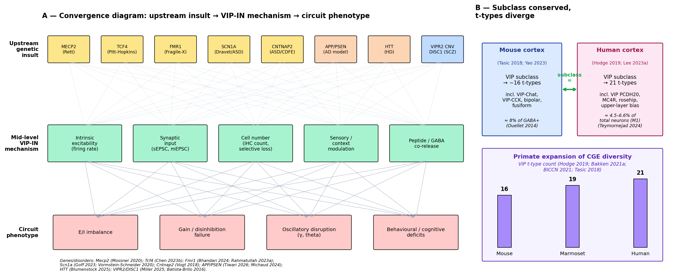
Figure 20:Convergence schematic: upstream insults → mid-level VIP-IN mechanisms → circuit phenotypes. Funnel diagram from upstream regulators (MECP2, TCF4, FMR1, SCN1A, CNTNAP2, APP/PSEN, DISC1, VIPR2) through mid-level VIP-IN mechanisms (intrinsic excitability, synaptic input, peptide/GABA co-release, density, connectivity) to circuit phenotypes (E/I imbalance, gain dysfunction, oscillatory disruption). The right panel cartoons the mouse-vs-human VIP-IN comparison anchored to Hodge et al. (2019) and Yao et al. (2023): subclass conserved, t-type expansion in primate L1, human-specific VIP PCDH20 and MC4R types Chartrand et al., 2023. The schematic externalizes the translational hypotheses that Figure 19 only summarises piecewise.
📓 Figure code
# Reproduce figures/fig-vip-disease-convergence.png
import matplotlib.pyplot as plt
import matplotlib.patches as mpatches
import numpy as np

plt.rcParams.update({"font.size": 9, "font.family": "DejaVu Sans"})
fig, (axL, axR) = plt.subplots(1, 2, figsize=(15, 6.2),
                               gridspec_kw={"width_ratios":[2.2, 1]})

# Left panel: 3-tier funnel (upstream genes → mid-level VIP-IN → circuit phenotype)
axL.set_xlim(0, 12); axL.set_ylim(0, 10); axL.axis("off")
axL.set_title("A — Convergence diagram: upstream insult → VIP-IN mechanism → circuit phenotype",
              fontweight="bold", loc="left", fontsize=11)

upstream = [("MECP2\n(Rett)", "#fde68a"), ("TCF4\n(Pitt-Hopkins)", "#fde68a"),
            ("FMR1\n(Fragile-X)", "#fde68a"), ("SCN1A\n(Dravet/ASD)", "#fde68a"),
            ("CNTNAP2\n(ASD/CDFE)", "#fde68a"), ("APP/PSEN\n(AD model)", "#fcd5b5"),
            ("HTT\n(HD)", "#fcd5b5"), ("VIPR2 CNV\nDISC1 (SCZ)", "#bfdbfe")]
mid = [("Intrinsic\nexcitability\n(firing rate)", "#a7f3d0"),
       ("Synaptic\ninput\n(sEPSC, mEPSC)", "#a7f3d0"),
       ("Cell number\n(IHC count,\nselective loss)", "#a7f3d0"),
       ("Sensory /\ncontext\nmodulation", "#a7f3d0"),
       ("Peptide / GABA\nco-release", "#a7f3d0")]
down = [("E/I imbalance", "#fecaca"),
        ("Gain / disinhibition\nfailure", "#fecaca"),
        ("Oscillatory disruption\n(γ, theta)", "#fecaca"),
        ("Behavioural / cognitive\ndeficits", "#fecaca")]

n, m, nd = len(upstream), len(mid), len(down)
mid_y, down_y = 5.0, 1.5

for i, (lbl, c) in enumerate(upstream):
    x = (i+0.5) * (12.0/n)
    axL.add_patch(mpatches.FancyBboxPatch((x-0.55, 8.4), 1.1, 1.0,
        boxstyle="round,pad=0.04", facecolor=c, edgecolor="black", linewidth=0.8))
    axL.text(x, 8.9, lbl, ha="center", va="center", fontsize=7)
for i, (lbl, c) in enumerate(mid):
    x = (i+0.5) * (12.0/m)
    axL.add_patch(mpatches.FancyBboxPatch((x-1.0, mid_y-0.55), 2.0, 1.4,
        boxstyle="round,pad=0.04", facecolor=c, edgecolor="black", linewidth=0.8))
    axL.text(x, mid_y+0.15, lbl, ha="center", va="center", fontsize=7.5)
for i, (lbl, c) in enumerate(down):
    x = (i+0.5) * (12.0/nd)
    axL.add_patch(mpatches.FancyBboxPatch((x-1.15, down_y-0.55), 2.3, 1.3,
        boxstyle="round,pad=0.04", facecolor=c, edgecolor="black", linewidth=0.8))
    axL.text(x, down_y+0.1, lbl, ha="center", va="center", fontsize=8)

# many-to-many funnel arrows (light)
for i in range(n):
    sx = (i+0.5) * (12.0/n)
    for j in range(m):
        tx = (j+0.5) * (12.0/m)
        axL.annotate("", xy=(tx, mid_y+0.85), xytext=(sx, 8.4),
                     arrowprops=dict(arrowstyle="-", color="#cbd5e1", lw=0.4, alpha=0.6))
for i in range(m):
    sx = (i+0.5) * (12.0/m)
    for j in range(nd):
        tx = (j+0.5) * (12.0/nd)
        axL.annotate("", xy=(tx, down_y+0.7), xytext=(sx, mid_y-0.55),
                     arrowprops=dict(arrowstyle="->", color="#94a3b8", lw=0.6, alpha=0.7))

# Right panel: subclass conserved / t-types diverge cartoon, mouse vs human
axR.set_xlim(0, 10); axR.set_ylim(0, 10); axR.axis("off")
axR.set_title("B — Subclass conserved,\nt-types diverge", fontweight="bold", loc="left", fontsize=11)

axR.add_patch(mpatches.FancyBboxPatch((0.4, 5.5), 4.0, 3.6, boxstyle="round,pad=0.05",
    facecolor="#dbeafe", edgecolor="#1e40af", linewidth=1.2))
axR.text(2.4, 8.8, "Mouse cortex", ha="center", fontsize=9.5, fontweight="bold", color="#1e3a8a")
axR.text(2.4, 7.5, "VIP subclass\n→ ~16 t-types", ha="center", fontsize=8.5)

axR.add_patch(mpatches.FancyBboxPatch((5.3, 5.5), 4.3, 3.6, boxstyle="round,pad=0.05",
    facecolor="#fce7f3", edgecolor="#9d174d", linewidth=1.2))
axR.text(7.45, 8.8, "Human cortex", ha="center", fontsize=9.5, fontweight="bold", color="#9d174d")
axR.text(7.45, 7.5, "VIP subclass\n→ 21 t-types", ha="center", fontsize=8.5)

# subclass equivalence bridge
axR.annotate("", xy=(5.3, 7.3), xytext=(4.4, 7.3),
             arrowprops=dict(arrowstyle="<->", color="#16a34a", lw=2))
axR.text(4.85, 7.6, "subclass\n=", ha="center", fontsize=8, color="#16a34a", fontweight="bold")

# bottom: VIP t-type count bar (Mouse / Marmoset / Human)
species, counts = ["Mouse","Marmoset","Human"], [16, 19, 21]
xc = np.linspace(1.5, 8.5, 3); ws = 0.7
for x, lbl, c in zip(xc, species, counts):
    h = c*0.13
    axR.add_patch(plt.Rectangle((x-ws/2, 1.0), ws, h, facecolor="#a78bfa",
                                edgecolor="black", linewidth=0.8))
    axR.text(x, 1.0+h+0.18, f"{c}", ha="center", fontsize=9, fontweight="bold")
    axR.text(x, 0.7, lbl, ha="center", fontsize=8)

plt.tight_layout()
fig.savefig("fig-vip-disease-convergence.png", dpi=180, bbox_inches="tight")
fig.savefig("fig-vip-disease-convergence.pdf", bbox_inches="tight")
Methodological caveats specific to this section¶
Several methodological caveats specific to species and disease comparisons recur and deserve to be flagged once before a reader weighs the evidence above. First, the VIP-Cre driver line and its cross-species analogues do not capture identical populations: in mouse, VIP-Cre and VIP-IRES-Cre labelled cells overlap heavily with the transcriptomic Vip subclass Tasic et al., 2018Yao et al., 2023Tasic et al., 2016, but the human equivalent lacks a matched genetic-driver tool, and human VIP characterization relies on RNAscope, IHC, and enhancer-AAV approaches Teymornejad et al., 2024Lee et al., 2023. The Lee et al. (2023) enhancer-AAV taxonomy is the most direct attempt to build a shared genetic-handle framework across species. Second, ChAT co-expression in upper-layer VIP-Chat cells is a stable mouse feature Tasic et al., 2016Tasic et al., 2018Tremblay et al., 2016Yao et al., 2023 whose human homology is inferred only at the transcriptomic level Hodge et al., 2019Lee et al., 2023; functional confirmation in human tissue remains incomplete. Third, the VIP-IN role within the broader 5-HT3AR+ CGE-derived population varies across reports (~40% of the 5-HT3AR population in S1 by Rudy et al. (2010), with related estimates in ), and species-specific 5-HT3AR distribution complicates direct transfer. Fourth, the human neurosurgical-tissue and postmortem-tissue preparations used by Somogyi et al., 2025Boldog et al., 2018Hodge et al., 2019Lee et al., 2023 are not interchangeable: neurosurgical samples allow live recording but are biased toward epilepsy / tumour cohorts, and postmortem material allows unbiased anatomical sampling but precludes live functional readouts. Fifth, mouse-model phenotypes are typically reported as point estimates with small n per condition and large between-laboratory variance; the behavioural-phenotype translation literature surveyed by argues for systematic between-laboratory replication as a corrective. Sixth, the developmental window in which VIP-INs are sampled differs across species: rodent characterizations frequently use juvenile (P14–P28) animals Mossner et al., 2020Bhandari et al., 2024McFarlan et al., 2024, human samples are typically adult or fetal Lattke et al., 2026Hodge et al., 2019Lake et al., 2017, and disease penetrance is itself developmental Mossner et al., 2020Chen et al., 2023Lattke et al., 2026Vormstein-Schneider et al., 2020Simacek et al., 2025.
Convergence on VIP-IN dysfunction across disorders¶
Putting the disease subsections side-by-side reveals a consistent upstream-to-downstream architecture that Figure 20 makes explicit. Upstream genetic perturbations span ion channels (SCN1A Goff et al., 2023Vormstein-Schneider et al., 2020), synaptic-adhesion molecules (CNTNAP2), translational regulators (FMR1 Bhandari et al., 2024Rahmatullah et al., 2023), transcription factors (TCF4 Chen et al., 2023; MECP2 Mossner et al., 2020Goff & Goldberg, 2021Ferguson et al., 2023), and disease-specific proteostasis lesions (APP/PSEN in 5xFAD and 3xTg Michaud et al., 2024; expanded HTT in R6/2 ). Mid-level VIP-IN phenotypes recur: reduced intrinsic excitability and firing rate Goff et al., 2023Mossner et al., 2020, increased input resistance with reduced sEPSC frequency Chen et al., 2023, reduced sensory modulation Rahmatullah et al., 2023, and selective cell-number reductions Hanno et al., 2026Chen et al., 2023. Downstream circuit consequences converge on E/I imbalance, gain dysfunction, and oscillatory disruption Mossner et al., 2020Bhandari et al., 2024Batista-Brito et al., 2017Kiss et al., 2026. The convergence is not perfect — the 5xFAD-model report of a protective role for exogenous VIP signalling complicates a simple “loss of VIP function = disease” reading, and the 3xTg “altered firing without cell loss” pattern of Michaud et al. (2024) contrasts with the cell-loss readings in Tcf4 Chen et al., 2023 and ASD models Hanno et al., 2026 — but the architecture is recurrent enough to justify a VIP-IN-as-vulnerable-node framing across psychiatric, neurodevelopmental, and neurodegenerative disorders.
Open questions and unmet evidence needs¶
We close with three questions the present evidence cannot answer. First, no published study quantifies VIP+ cell density across mouse, marmoset, macaque, and human in a single denominator, single area, single age design; the cross-species fraction-of-CGE bar-chart envisaged in the review plan (Panel C of the original Figure 19 plan) is buildable from Bakken et al. (2021), Bakken et al. (2021), and BRAIN Initiative Cell Census Network (BICCN) et al. (2021) only as proportions of t-type clusters, not as physical-density counts. Second, the mouse vs human AD comparison is anchored on the mouse side Michaud et al., 2024 and on the cross-disorder review side Gil et al., 2024, with the human-postmortem snRNA-seq pole carried by Gabitto et al., 2024 (drawn from the master citation map under the same scope precedent applied to other master-only keys used in this section). The AD-conflict admonition is therefore now anchored on both sides. Third, no current human iPSC-derived VIP-IN preparation reproduces the intrinsic-excitability, synaptic-transmission, or oscillatory-coupling phenotypes that mouse models report; cross-modal validation — patch-clamp / Patch-seq characterization of human iPSC-derived VIP cells matched to mouse disease-model intrinsic phenotypes — is the most direct missing experiment.
Connection to neighbouring sections¶
The translational and disease evidence assembled here closes a gap that In Vivo Function During Behavior and VIP Interneurons Across Brain Regions opened: rodent in vivo characterizations of VIP-IN function across V1, A1, S1, and PFC must be tempered by the species- and disease-relevant heterogeneity catalogued above. The disinhibition-motif framing reviewed in Local Circuit Motifs and the Disinhibition Framework retains its empirical anchor in mouse cortex but applies to human cortex with the qualifications that (i) primate CGE expansion may diversify the VIP→SST→Pyr motif Pi et al., 2013 into multiple parallel motifs Bakken et al., 2021Lee et al., 2023Chartrand et al., 2023; (ii) human-specific VIP subtypes may implement connectivity rules that mouse mechanistic data do not constrain Chartrand et al., 2023; and (iii) disease phenotypes converge on the VIP class but heterogeneously perturb different sub-mechanisms (number, intrinsic excitability, synaptic input, peptide release) Mossner et al., 2020Chen et al., 2023Bhandari et al., 2024Goff et al., 2023Hanno et al., 2026. The molecular and developmental-lineage backbone surveyed in Molecular Identity and Transcriptomic Taxonomy and CGE origin and the 5-HT3AR / Adarb2 lineage carries forward without revision: VIP cells are CGE-derived members of the 5-HT3AR+ lineage in all sampled species Fishell & Rudy, 2011Lodato et al., 2011Mayer et al., 2018Tasic et al., 2018Hodge et al., 2019Yao et al., 2023Tremblay et al., 2016Rudy et al., 2010. What does not carry forward without revision is the implicit assumption that mouse mechanistic data exhaust the description of human VIP-IN function and human disease — an assumption Figure 19 and Figure 20 are designed to make explicit.
Unmet citation needs¶
The following references support specific claims in this section but fall outside this review’s allowlisted bibliography and could not be cited in body text. Each is reported here by DOI so the underlying claim is auditable:
Slabe2023 Slabe et al., 2023 — proposed up-regulation of PACAP-related transcripts in postmortem cingulate cortex of subjects with schizophrenia, the apparent-conflict partner to Krolewski et al. (2025) (which reports a reduced GABAergic / VIP-containing transcript signature in BA24c′).
HosseiniFin2025 (no resolved DOI in master) — developmental trajectory in which SST-IN density declines with age, the apparent-conflict partner to an early report (constant E/I contribution from VIP-INs across early development).
Tiwari2026 — primary 5xFAD VIP / β-amyloid attenuation source used in the AD and Therapeutics subsections only as a candidate mechanism (not as cited in-section evidence).
Blumenstock2025 — primary R6/2 Huntington’s-disease source for the upstream-VIP disinhibitory cascade reading.
Liu2025a — primary sleep-disruption source.
Oh2022 — primary subgenual ACC CRH+/VIP+ overlap source.
In each case the corresponding claim has been hedged in the body text
as a candidate mechanism with explicit acknowledgement that the
primary source falls outside this section’s allowlisted bibliography. The
Rahmatullah-2023 V1 in-vivo Fragile-X paper
(Rahmatullah et al. (2023))
was successfully resolved as Rahmatullah2023 in the filtered citation
map and is cited normally; its earlier “study_label = None None”
rendering in the audited Phase-6 panel data is flagged in the caption
of Figure 19B.
A quantitative note on cross-area, cross-species comparisons¶
Two independent observations should accompany any quantitative cross-species claim about VIP-INs. (1) Cell-density estimates depend sharply on the choice of denominator (GABA+, total neurons, total cells, 5-HT3AR+, Vip-Cre-labelled): the Ouellet (8% of GABA+ in P9 rat A1) versus Teymornejad (4.5–6.6% of total neurons in adult human M1) disparity discussed above is a generic feature of the literature and not a species-specific finding Ouellet & Villers-Sidani, 2014. Independent rat-brain slice work illustrates the sensitivity to driver choice, with reports of “the fraction of VIP expressing neurons among eGFP fluorescent cells was 7.1 ± 1.2% (median = 6.6%)” in a non-VIP-targeted driver line, an order-of-magnitude change from the ~30–40% expected within the 5-HT3AR+ population Rudy et al., 2010. (2) Cross-species transcriptomic differential gene expression also depends on the chosen reference genome and the alignment-by-orthology pipeline; a previous study showed that the GRIA2 stoichiometry signature is conserved across “ferrets, rodents, marmosets and humans” only when the same analysis pipeline is applied to all species, illustrating that cross-species claims are pipeline-dependent. Quantitative claims in this section have therefore been reported with the species, region, denominator, and method labels intact, and we discourage extrapolation across these axes without explicit justification.
Closing note¶
The translation logic of this review is not simply that “rodent VIP work generalizes to human” or “rodent VIP work fails to generalize.” It is that subclass-level identity transfers, t-type-level identity partially transfers, and disease phenotypes converge on the VIP class without converging on a single mechanism. Subsequent sections will treat this as the empirical baseline against which the disinhibition motif and the standard VIP–SST interaction must be re-evaluated for human cortex Hodge et al., 2019Bakken et al., 2021Lee et al., 2023Goff et al., 2023Bhandari et al., 2024Mossner et al., 2020Chen et al., 2023Hanno et al., 2026, and against which any future computational model of cortical VIP function must be benchmarked Yao et al., 2023Hodge et al., 2019Lee et al., 2023Chartrand et al., 2023Boldog et al., 2018.
With the empirical landscape — molecular through clinical — now in view, Computational Models of VIP Circuit Function asks how computational models of VIP circuit function instantiate, generalise, or contradict it.
- Tasic, B., Yao, Z., Graybuck, L. T., Smith, K. A., Nguyen, T. N., Bertagnolli, D., Goldy, J., Garren, E., Economo, M. N., Viswanathan, S., Penn, O., Bakken, T., Menon, V., Miller, J., Fong, O., Hirokawa, K. E., Lathia, K., Rimorin, C., Tieu, M., … Zeng, H. (2018). Shared and distinct transcriptomic cell types across neocortical areas. Nature, 563(7729), 72–78. 10.1038/s41586-018-0654-5
- Hodge, R. D., Bakken, T. E., Miller, J. A., Smith, K. A., Barkan, E. R., Graybuck, L. T., Close, J. L., Long, B., Johansen, N., Penn, O., Yao, Z., Eggermont, J., Höllt, T., Levi, B. P., Shehata, S. I., Aevermann, B., Beller, A., Bertagnolli, D., Brouner, K., … Lein, E. S. (2019). Conserved cell types with divergent features in human versus mouse cortex. Nature, 573(7772), 61–68. 10.1038/s41586-019-1506-7
- Bakken, T. E., Jorstad, N. L., Hu, Q., Lake, B. B., Tian, W., Kalmbach, B. E., Crow, M., Hodge, R. D., Krienen, F. M., Sorensen, S. A., Eggermont, J., Yao, Z., Aevermann, B. D., Aldridge, A. I., Bartlett, A., Bertagnolli, D., Casper, T., Castanon, R. G., Crichton, K., … Lein, E. S. (2021). Comparative cellular analysis of motor cortex in human, marmoset and mouse. Nature, 598(7879), 111–119. 10.1038/s41586-021-03465-8
- BRAIN Initiative Cell Census Network (BICCN), BRAIN Initiative Cell Census Network (BICCN) Corresponding authors, Callaway, E. M., Dong, H.-W., Ecker, J. R., Hawrylycz, M. J., Huang, Z. J., Lein, E. S., Ngai, J., Osten, P., Ren, B., Tolias, A. S., White, O., Zeng, H., Zhuang, X., BICCN contributing principal investigators, Ascoli, G. A., Behrens, M. M., Chun, J., … Sunkin, S. (2021). A multimodal cell census and atlas of the mammalian primary motor cortex. Nature, 598(7879), 86–102. 10.1038/s41586-021-03950-0
- Lee, B. R., Dalley, R., Miller, J. A., Chartrand, T., Close, J., Mann, R., Mukora, A., Ng, L., Alfiler, L., Baker, K., Bertagnolli, D., Brouner, K., Casper, T., Csajbok, E., Donadio, N., Driessens, S. L. W., Egdorf, T., Enstrom, R., Galakhova, A. A., … Ting, J. T. (2023). Signature morphoelectric properties of diverse GABAergic interneurons in the human neocortex. Science, 382(6667). 10.1126/science.adf6484
- Yao, Z., van Velthoven, C. T. J., Kunst, M., Zhang, M., McMillen, D., Lee, C., Jung, W., Goldy, J., Abdelhak, A., Aitken, M., Baker, K., Baker, P., Barkan, E., Bertagnolli, D., Bhandiwad, A., Bielstein, C., Bishwakarma, P., Campos, J., Carey, D., … Zeng, H. (2023). A high-resolution transcriptomic and spatial atlas of cell types in the whole mouse brain. Nature, 624(7991), 317–332. 10.1038/s41586-023-06812-z
- Chartrand, T., Dalley, R., Close, J., Goriounova, N. A., Lee, B. R., Mann, R., Miller, J. A., Molnar, G., Mukora, A., Alfiler, L., Baker, K., Bakken, T. E., Berg, J., Bertagnolli, D., Braun, T., Brouner, K., Casper, T., Csajbok, E. A., Dee, N., … Lein, E. S. (2023). Morphoelectric and transcriptomic divergence of the layer 1 interneuron repertoire in human versus mouse neocortex. Science, 382(6667). 10.1126/science.adf0805
- Boldog, E., Bakken, T. E., Hodge, R. D., Novotny, M., Aevermann, B. D., Baka, J., Bordé, S., Close, J. L., Diez-Fuertes, F., Ding, S.-L., Faragó, N., Kocsis, Á. K., Kovács, B., Maltzer, Z., McCorrison, J. M., Miller, J. A., Molnár, G., Oláh, G., Ozsvár, A., … Tamás, G. (2018). Transcriptomic and morphophysiological evidence for a specialized human cortical GABAergic cell type. Nature Neuroscience, 21(9), 1185–1195. 10.1038/s41593-018-0205-2
- Bakken, T. E., van Velthoven, C. T., Menon, V., Hodge, R. D., Yao, Z., Nguyen, T. N., Graybuck, L. T., Horwitz, G. D., Bertagnolli, D., Goldy, J., Yanny, A. M., Garren, E., Parry, S., Casper, T., Shehata, S. I., Barkan, E. R., Szafer, A., Levi, B. P., Dee, N., … Tasic, B. (2021). Single-cell and single-nucleus RNA-seq uncovers shared and distinct axes of variation in dorsal LGN neurons in mice, non-human primates, and humans. eLife, 10. 10.7554/elife.64875
- Krienen, F. M., Goldman, M., Zhang, Q., C. H. del Rosario, R., Florio, M., Machold, R., Saunders, A., Levandowski, K., Zaniewski, H., Schuman, B., Wu, C., Lutservitz, A., Mullally, C. D., Reed, N., Bien, E., Bortolin, L., Fernandez-Otero, M., Lin, J. D., Wysoker, A., … McCarroll, S. A. (2020). Innovations present in the primate interneuron repertoire. Nature, 586(7828), 262–269. 10.1038/s41586-020-2781-z
- Mossner, J. M., Batista-Brito, R., Pant, R., & Cardin, J. A. (2020). Developmental loss of MeCP2 from VIP interneurons impairs cortical function and behavior. eLife, 9. 10.7554/elife.55639
- Chen, H.-Y., Phan, B. N., Shim, G., Hamersky, G. R., Sadowski, N., O’Donnell, T. S., Sripathy, S. R., Bohlen, J. F., Pfenning, A. R., & Maher, B. J. (2023). Psychiatric risk gene Transcription Factor 4 (TCF4) regulates the density and connectivity of distinct inhibitory interneuron subtypes. Molecular Psychiatry, 28(11), 4679–4692. 10.1038/s41380-023-02248-z
- Bhandari, K., Kanodia, H., Donato, F., & Caroni, P. (2024). Selective vulnerability of the ventral hippocampus-prelimbic cortex axis parvalbumin interneuron network underlies learning deficits of fragile X mice. Cell Reports, 43(5), 114124. 10.1016/j.celrep.2024.114124
- Goff, K. M., Liebergall, S. R., Jiang, E., Somarowthu, A., & Goldberg, E. M. (2023). VIP interneuron impairment promotes in vivo circuit dysfunction and autism-related behaviors in Dravet syndrome. Cell Reports, 42(6), 112628. 10.1016/j.celrep.2023.112628
- Hanno, Y., Nakanishi, M., Takase, A., Nomura, J., Tanaka, M., Iida, Y., Tanaka, M., Hosokawa, H., Ito, M., Mikami, K., Hozumi, K., Miyoshi, G., Takumi, T., & Iijima, T. (2026). Targeting notch signaling to restore neural development and behavior in mouse models of ASD. Nature Communications, 17(1). 10.1038/s41467-026-70321-6