Sections Molecular Identity and Transcriptomic Taxonomy and Developmental Origins and Postnatal Maturation established the molecular and developmental scaffold of the VIP-expressing class; this section asks how those categories project into morphological space. The contemporary picture, drawn from Patch-seq atlases, large-volume connectomics, and decades of biocytin-based reconstructions, is consistent on the gross descriptors but contested on the boundaries: cortical VIP cells span a recognisable but graded morphological space dominated by bipolar/bitufted somatodendritic profiles with descending axonal arbors Apicella & Marchionni, 2022, with smaller proportions of small-basket and arcade-like variants Rudy et al., 2010Yayon et al., 2022. Subclass-level (VIP vs SST vs PV) morphological separation is robust in cross-modal taxonomies Gouwens et al., 2019Gouwens et al., 2020Gliko et al., 2024Elabbady et al., 2025, while within-VIP clustering forms a continuum punctuated by reproducible centroids whose proportions vary across cortical area, layer, and species Gouwens et al., 2020Gouwens et al., 2020Prönneke et al., 2015Chartrand et al., 2023.
Canonical morphological types
The earliest single-cell descriptions of cortical VIP-expressing interneurons recognised a small set of recurring forms. Kawaguchi & Kubota (1996) described four morphological classes in rat frontal cortex — vertically bipolar cells with predominantly descending axonal collaterals, double-bouquet cells, small-basket cells with short and dense local arbors, and arcade-shaped cells with arching axonal trees — and showed that VIP-immunoreactive axons terminated in symmetrical (inhibitory) synapses with a strong vertical bias Kawaguchi & Kubota, 1996Cauli et al., 1997Cauli et al., 2014Apicella & Marchionni, 2022. Cauli et al. (1997) extended the framework by combining whole-cell recording with single-cell RT-PCR in rat sensorimotor cortex, identifying a vertically oriented bipolar subset that co-expressed calretinin and VIP and fired in an irregular-spiking pattern Cauli et al., 1997Cauli et al., 2014Vucurovic et al., 2010Stachniak et al., 2021. Vucurovic et al. (2010) then demonstrated, using 5-HT3aR-BAC transgenics combined with in situ hybridisation, that the cortical 5-HT3AR-positive lineage partitions into a VIP-positive subset with bipolar/bitufted morphology and a VIP-negative subset with multipolar morphology — embedding the bipolar/bitufted descriptor in the broader CGE taxonomy Vucurovic et al., 2010Rudy et al., 2010Cauli et al., 2014Fishell & Kepecs, 2020.
The bipolar/bitufted morphotype dominates in numerical surveys but does not exhaust the class. Reviews of the rodent literature converge on a list of bipolar, bitufted, multipolar, small-basket, and arcade morphologies, with VIP-positive basket cells representing a numerical minority of the class Rudy et al., 2010Cauli et al., 2014Apicella & Marchionni, 2022Fishell & Kepecs, 2020. An earlier report, using VIP-Cre × tdTomato barrel cortex slices, reported a substantially broader repertoire than the classical bipolar form — including bipolar/bitufted, tufted, and multipolar somatodendritic configurations distributed differently across layers — and noted that the major axon often emerged not from the soma but from a basal dendrite directed toward the white matter, a feature also flagged by an earlier report Kawaguchi & Kubota, 1996. Two intersectional studies further partitioned the class along molecular axes: Stachniak et al. (2021) separated CR-positive bipolar and CCK-positive multipolar VIP subtypes during cortical development, and Yayon et al. (2022) resolved the cholinergic subset (VChIs) of VIP barrel-cortex cells into bipolar (two stem dendrites) and multipolar (≥3 stem dendrites) morphologies Stachniak et al., 2021Yayon et al., 2022Wong et al., 2022Prönneke et al., 2015.
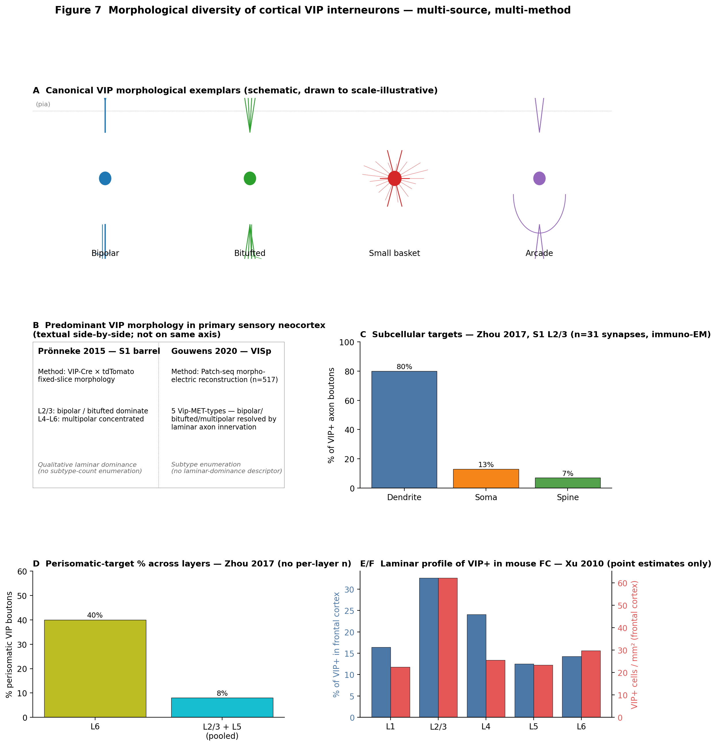
Figure 5:Morphological diversity of cortical VIP interneurons. (A) Idealised exemplar morphologies — bipolar, bitufted, small-basket, and arcade — drawn to illustrate the canonical VIP morphological repertoire described by earlier studies and Cauli et al. (2014). (B) Textual side-by-side of predominant VIP morphological subtype reported in two genetically defined VIP-Cre datasets in mouse primary sensory neocortex: a preceding study (S1 barrel) versus Gouwens et al. (2020) (VISp). Caveat (verbatim, Phase 6 audit): Render as textual side-by-side, not as a quantitative bar (per restructure_notes). | State explicitly that Pronneke reports laminar dominance in S1 barrel cortex while Gouwens reports subtype enumeration across Patch-seq met-types in VISp — entries are not on the same axis. | Note difference in cortical area (S1 vs VISp) and method (fixed-slice morphology vs Patch-seq morpho-electric). (C) Subcellular target distribution of VIP-axon boutons in mouse S1 barrel L2/3 (immuno-EM, n=31 synapses) from an initial investigation. Caveat (verbatim, Phase 6 audit): Specify the sample size (n=31 synapses, L2/3) directly in the panel. | Verify in the published Methods/Results that 80/13/7 are L2/3-specific values (not the cross-laminar overall reported in abstract). If unverifiable, label the row as ‘all layers (Zhou 2017)’ rather than L2/3. (D) Within-paper layer comparison of VIP-bouton perisomatic-target percentage (L6 vs L2/3+L5 pooled) from Zhou et al. (2017). Caveat (verbatim, Phase 6 audit): State that per-layer n_synapses denominators were not extractable; bars show point estimates without error. | Indicate that one row is a single layer (L6) while the other is pooled across L2/3 and L5. | Source: Zhou 2017 (10.1093/cercor/bhx220), serial-section EM in mouse S1 barrel cortex. (E) Laminar percent-of-VIP+ profile in mouse frontal cortex from Xu et al. (2009). Caveat (verbatim, Phase 6 audit): Single-study ‘comparison’ — splitter yields a single laminar profile; no cross-paper comparison possible. | No value_source_sentence quoted for the layer percentages (verifiability gap). | No per-layer n provided; sample_size flagged in restructure_notes. (F) Laminar density (cells/mm²) of VIP+ neurons in mouse frontal cortex from Xu et al. (2009). Caveat (verbatim, Phase 6 audit): State that n_definition (animals/sections) and dispersion are not reported in the source extraction; values shown are point estimates only. | Indicate the source is Xu 2010 (10.1002/cne.22229), immunohistochemistry/cell-counting in mouse frontal cortex. | If the paper’s Tables report frontal cortex separately, cite the specific table; otherwise note the regional attribution caveat.
📓 Figure code
import matplotlib.pyplot as plt
import numpy as np
from matplotlib.patches import Rectangle, Circle

fig = plt.figure(figsize=(13, 14))
gs = fig.add_gridspec(3, 2, hspace=0.55, wspace=0.30, height_ratios=[1.1, 1.0, 1.0])

# Panel A — exemplar morphologies (drawn schematic)
axA = fig.add_subplot(gs[0, :])
axA.set_xlim(0,4); axA.set_ylim(0,1); axA.axis("off")
axA.set_title("A  Canonical VIP morphological exemplars (schematic)", loc="left", fontsize=11, fontweight="bold")

def draw_bipolar(ax, cx, cy, color="#1f77b4"):
    ax.plot([cx, cx], [cy+0.29, cy+0.64], color=color, lw=1.6)
    ax.plot([cx, cx], [cy-0.29, cy-0.64], color=color, lw=1.6)
    ax.plot([cx-0.02, cx-0.02], [cy-0.29, cy-1.0], color=color, lw=0.9)
    ax.add_patch(Circle((cx, cy), 0.04, color=color))

def draw_bitufted(ax, cx, cy, color="#2ca02c"):
    for sign in (1, -1):
        for dx in [-0.06, -0.02, 0.02, 0.06]:
            ax.plot([cx, cx+dx], [cy+sign*0.29, cy+sign*0.64], color=color, lw=1.0)
    ax.plot([cx+0.01, cx+0.01], [cy-0.29, cy-0.96], color=color, lw=0.9)
    ax.add_patch(Circle((cx, cy), 0.04, color=color))

def draw_smallbasket(ax, cx, cy, color="#d62728"):
    for ang in np.linspace(0, 2*np.pi, 7)[:-1]:
        ax.plot([cx, cx+0.10*np.cos(ang)], [cy, cy+0.20*np.sin(ang)], color=color, lw=1.0)
    ax.add_patch(Circle((cx, cy), 0.045, color=color))

def draw_arcade(ax, cx, cy, color="#9467bd"):
    for sign in (1, -1):
        for dx in [-0.04, 0.04]:
            ax.plot([cx, cx+dx], [cy+sign*0.29, cy+sign*0.58], color=color, lw=1.0)
    th = np.linspace(np.pi, 2*np.pi, 60)
    ax.plot(cx + 0.18*np.cos(th), cy - 0.10 + 0.30*np.sin(th)*0.8, color=color, lw=0.9)
    ax.add_patch(Circle((cx, cy), 0.04, color=color))

xs = [0.5, 1.5, 2.5, 3.5]
for x, lab, dfn in zip(xs, ["Bipolar","Bitufted","Small basket","Arcade"],
                       [draw_bipolar, draw_bitufted, draw_smallbasket, draw_arcade]):
    dfn(axA, x, 0.5)
    axA.text(x, 0.02, lab, ha="center", fontsize=10)

# Panel B — Pronneke vs Gouwens textual side-by-side
axB = fig.add_subplot(gs[1, 0]); axB.axis("off")
axB.set_title("B  Predominant VIP morphology — textual side-by-side", loc="left", fontsize=10.5, fontweight="bold")
axB.text(0.02, 0.92, "Pronneke 2015 (S1 barrel)\nVIP-Cre fixed slice\nL2/3: bipolar/bitufted dominate\nL4-L6: multipolar concentrated", fontsize=9, va="top")
axB.text(0.55, 0.92, "Gouwens 2020 (VISp)\nPatch-seq, n=517\n5 Vip-MET-types resolved\nby laminar axon innervation", fontsize=9, va="top")

# Panel C — Zhou 2017 L2/3 subcellular targets (n=31)
axC = fig.add_subplot(gs[1, 1])
axC.bar(["Dendrite","Soma","Spine"], [80, 13, 7],
        color=["#4c78a8","#f58518","#54a24b"], edgecolor="black", lw=0.5)
for i, v in enumerate([80,13,7]):
    axC.text(i, v+1.5, f"{v}%", ha="center", fontsize=9)
axC.set_ylabel("% of VIP+ axon boutons"); axC.set_ylim(0, 100)
axC.set_title("C  Subcellular targets — Zhou 2017 (n=31, L2/3, immuno-EM)", loc="left", fontsize=10.5, fontweight="bold")

# Panel D — Zhou perisomatic-by-layer
axD = fig.add_subplot(gs[2, 0])
axD.bar(["L6", "L2/3+L5"], [40, 8], color=["#bcbd22","#17becf"], edgecolor="black", lw=0.5)
for i, v in enumerate([40, 8]):
    axD.text(i, v+1, f"{v}%", ha="center", fontsize=9)
axD.set_ylabel("% perisomatic VIP boutons"); axD.set_ylim(0, 60)
axD.set_title("D  Perisomatic-target % across layers — Zhou 2017", loc="left", fontsize=10.5, fontweight="bold")

# Panel E/F — Xu 2010 frontal cortex laminar profile
axE = fig.add_subplot(gs[2, 1])
layers = ["L1","L2/3","L4","L5","L6"]
pcts = [16.4, 32.6, 24.1, 12.5, 14.3]
dens = [22.4, 62.1, 25.5, 23.4, 29.8]
x = np.arange(len(layers)); w = 0.4
axE.bar(x-w/2, pcts, w, color="#4c78a8")
axE2 = axE.twinx()
axE2.bar(x+w/2, dens, w, color="#e45756")
axE.set_xticks(x); axE.set_xticklabels(layers)
axE.set_ylabel("% of VIP+ in FC", color="#4c78a8")
axE2.set_ylabel("VIP+ cells / mm^2 (FC)", color="#e45756")
axE.set_title("E/F  Laminar profile — Xu 2010 (point estimates only)", loc="left", fontsize=10.5, fontweight="bold")

fig.suptitle("Figure 7  Morphological diversity of cortical VIP interneurons", fontsize=12.5, y=0.995, fontweight="bold")
fig.savefig("fig-vip-morphologies.png", dpi=200, bbox_inches="tight")
fig.savefig("fig-vip-morphologies.pdf", bbox_inches="tight")
plt.close(fig)
Subtype proportions and laminar distribution
Numerical estimates of how the canonical morphologies partition the VIP class differ substantially across studies, reflecting genuine biology and methodological choice. Gonchar (2008) reported that VIP-expressing GABAergic neurons account for roughly 11% of cortical interneurons in mouse V1 and are concentrated in supragranular layers, with CR-, VIP-, ChAT- and CCK-immunoreactive populations enriched in L2/3 Gonchar, 2008Xu et al., 2009Rudy et al., 2010Cauli et al., 2014. Xu et al. (2009) quantified absolute laminar densities across mouse frontal cortex, somatosensory cortex, and V1, finding L2/3 VIP-positive density 2–3× greater than deeper layers (e.g. ~62 cells/mm² in L2/3 versus ~22–30 in other layers in frontal cortex, with 32.6% of all VIP-positive cells in L2/3) Xu et al., 2009Gonchar, 2008Prönneke et al., 2015Apicella & Marchionni, 2022. The Allen Institute Patch-seq atlas Gouwens et al., 2020Gouwens et al., 2019Gliko et al., 2024Gouwens et al., 2020 partitioned the VISp Vip subclass into five morpho-electric-transcriptomic types (Vip-MET-1 through Vip-MET-5) all resembling bipolar or bitufted cells but differing in laminar axon innervation, with Vip-MET-5 (Vip Crispld2) the principal L1-projecting type and Vip-MET-1/-2 occupying L2/3–L4 with distinct axonal biases.
The proportions disagree where the population definitions and methods diverge. The classical estimate that approximately 40% of cortical VIP interneurons co-express calretinin Rudy et al., 2010Cauli et al., 2014Cauli et al., 1997De Marco García et al., 2011 was challenged by region-specific counts in mouse V1 layers 4 and 5 reporting ~52% VIP/CR overlap Gonchar, 2008Rudy et al., 2010Cauli et al., 2014.
Other proportional estimates extend rather than overturn this picture. De Marco García et al. (2011) separated VIP-positive multipolar cells from CR-positive bipolar/multipolar siblings in early postnatal cortex, with the two showing distinct activity-dependence of axonal arbor length under in utero electroporation De Marco García et al., 2011Stachniak et al., 2021Wong et al., 2022Fishell & Kepecs, 2020. Wong et al. (2022) reported, using immunohistochemistry of CGE-derived Htr3a-positive cells, that VIP is expressed in two morphologically distinct subtypes — most CR-positive bipolar cells and approximately half of CR-/Reln-negative basket cells — providing a molecular link between the bipolar majority and the smaller VIP-basket fraction Wong et al., 2022Stachniak et al., 2021Rudy et al., 2010Yayon et al., 2022. Emmenegger et al. (2018) confirmed that intra-columnar VIP-like cells with bipolar dendrites form a distinct minority of L4 non-fast-spiking types in rat barrel cortex Emmenegger et al., 2018Prönneke et al., 2015Cauli et al., 1997Apicella & Marchionni, 2022.
Discrete subtypes versus a graded continuum
Whether within-VIP variation is best modelled as discrete cell types or as continuous gradients is a recurring methodological question. The Allen Institute Patch-seq programme has produced the most extensive substrate for this question. Gouwens et al. (2019) first reported that Patch-clamp/biocytin morphology in adult mouse VISp supports a taxonomy of ~17 e-types, 38 m-types and 46 me-types, with axonal (rather than dendritic) features the dominant discriminator and good correspondence to transcriptomic subclasses including VIP Gouwens et al., 2019Gouwens et al., 2020Gliko et al., 2024Schneider-Mizell et al., 2025. Gouwens et al. (2020) extended this to morpho-electric-transcriptomic met-types and resolved five Vip-MET clusters, while a companion preprint Gouwens et al., 2020Gouwens et al., 2019Gliko et al., 2024Prönneke et al., 2015 cautioned that >3,700 mouse VISp Patch-seq interneurons (350 reconstructed) reveal substantial continuous morpho-electric variation between and within met-types. Gliko et al. (2024) then enlarged the reconstructed inhibitory dataset to 813 cells and showed that arbor-density representations alone classify subclass-level identity (Sst/Pvalb/Vip/Lamp5) with high accuracy while within-VIP boundaries remain less crisp Gliko et al., 2024Gouwens et al., 2020Gouwens et al., 2020Elabbady et al., 2025.
Connectomic reconstructions converge on the same picture from a complementary direction. Schneider-Mizell et al. (2025) identified, in the MICrONS cubic-millimetre EM volume of mouse VISp, an “inhibition-targeting” cell class (InhTCs) that aligns with the disinhibitory VIP cardinal class and that subdivides into two connectomic subtypes — InhTC_dist (targeting distal-dendrite SST-like cells) and InhTC_peri (targeting perisomatic PV-like basket cells) — refining the long-standing view of a single VIP→SST→Pyr motif Schneider-Mizell et al., 2025Lee et al., 2013Gamlin et al., 2025Cauli et al., 2014. Elabbady et al. (2025) showed that perisomatic ultrastructural features alone classify mouse-cortex inhibitory neurons into bipolar (VIP-overlapping), basket, Martinotti, and neurogliaform classes; in the same volume, 6,805 expert-classified inhibitory cells included 997 bipolar cells (~14.6%) operationally defined by 2–3 primary dendrites and synapses primarily onto other inhibitory neurons Elabbady et al., 2025Schneider-Mizell et al., 2025Gouwens et al., 2020Gliko et al., 2024. Gamlin et al. (2025) used Patch-seq–EM joint reconstructions to confirm that Vip cells exhibit a stereotyped narrow-horizontal/long-vertical axon arbor, with 20/22 EM-reconstructed Vip cells (≈91%) preferentially targeting inhibitory cells Gamlin et al., 2025Schneider-Mizell et al., 2025Elabbady et al., 2025Lee et al., 2013.
Axonal architecture and target compartment selection
The bipolar/bitufted somatodendritic profile is matched on the axonal side by a vertically biased, mostly inhibition-targeting arbor. Kawaguchi & Kubota (1996)’s original report that VIP-immunoreactive axons terminated in symmetrical synapses with a vertical (radially oriented) bias has been recovered repeatedly in transgenic and viral preparations Kawaguchi & Kubota, 1996Cauli et al., 2014Apicella & Marchionni, 2022Prönneke et al., 2015. The most quantitative compartment-level data come from Zhou et al. (2017), who used immuno-electron microscopy in mouse barrel cortex to show that VIP boutons innervate primarily dendrites (~80% of synapses, n=31 in L2/3), with somata (13%) and spines (7%) sparsely targeted in upper layers Zhou et al., 2017Lee et al., 2013Gulyás et al., 1996Apicella & Marchionni, 2022. The same study reported a striking layer-dependent shift: in L6, ~40% of VIP boutons synapsed onto somata, contrasting with 8% in L2/3 and 8% in L5 — indicating that the canonical “perisomatic basket cell” function is not absent from the VIP class but compartmentalised into deep layers, and aligning with the layer-specific axon innervation that defines Patch-seq met-types Gouwens et al., 2020Schneider-Mizell et al., 2025Gamlin et al., 2025.
A second, more contested feature concerns the origin of the major axon. A preceding study reported that the majority of VIP barrel-cortex cells emit their main axon not from the soma but from a basal dendrite directed toward the white matter — a feature shared across the dataset and corroborated by an early report and consistent with reconstructions reviewed by a previous study Kawaguchi & Kubota, 1996. Outside primary sensory neocortex, the picture is less clear: in medial entorhinal cortex (MEC), Badrinarayanan et al. (2021) recorded 60 VIP cells (17 reconstructed) and reported bitufted or multipolar morphologies with layer-specific axonal asymmetry — superficial cells extended axons into deep layers, deep cells made almost no contact with superficial layers — but did not specifically report the dendrite-emerging-axon majority described in neocortex Badrinarayanan et al., 2021Apicella & Marchionni, 2022.
The interneuron-targeting bias is itself a defining structural property. Gulyás et al. (1996) first showed in rat hippocampus that calretinin-immunoreactive interneurons (homologous to cortical CR/VIP interneuron-selective cells) preferentially target other GABAergic interneurons — including VIP-containing basket cells — while sparing pyramidal neurons Gulyás et al., 1996Lee et al., 2013Schneider-Mizell et al., 2025Gamlin et al., 2025. Lee et al. (2013) extended this to neocortex with channelrhodopsin-assisted circuit mapping in mouse barrel cortex, showing that VIP cells preferentially inhibit SST-expressing interneurons during whisking Lee et al., 2013Gulyás et al., 1996Schneider-Mizell et al., 2025Prönneke et al., 2015. The MICrONS connectome converts this into an EM-grounded statistic: across the volume, VIP-aligned bipolar/InhTC cells make most of their synapses onto inhibitory targets, with InhTC_dist preferentially contacting SST-like distal-dendrite cells and InhTC_peri preferentially contacting PV-like basket cells Schneider-Mizell et al., 2025Elabbady et al., 2025Gamlin et al., 2025Lee et al., 2013.
Patch-seq met-types and the morpho-electric structure within VIP
Resolution within the VIP class has tightened around the five Patch-seq met-types defined in mouse VISp. Gouwens et al. (2020) reported that all five Vip-MET-types resemble bipolar or bitufted cells but differ in laminar axon innervation: Vip-MET-5 (containing Vip Crispld2 t-types) is the principal L1-projecting VIP met-type, innervates all cortical layers including L1, and shows elevated hyperpolarisation-evoked sag relative to the other Vip met-types Gouwens et al., 2020Gouwens et al., 2019Chartrand et al., 2023. Vip-MET-1 occupies L2/3–L4 with bitufted dendrites extending into L1 and a concentrated axon plexus in lower L2/3, while Vip-MET-2 shares the laminar position of Vip-MET-1 but exhibits a deep-biased and broader axon distribution — making the laminar axon profile the defining within-VIP discriminator rather than dendritic geometry alone Gouwens et al., 2020Gouwens et al., 2019Schneider-Mizell et al., 2025Gamlin et al., 2025. Gouwens et al. (2019) had earlier shown across the whole inhibitory inventory that the axon, not the dendrite, is the dominant feature distinguishing inhibitory m-types in mouse visual cortex; the same paper noted that L2/3 Chat-Cre-labelled neurons (a known subset of Vip cells) map predominantly to a small number of related me-types associated with the Vip-Ptprt-Pkp2 transcriptomic type Gouwens et al., 2019Gouwens et al., 2020Yayon et al., 2022Wong et al., 2022.
The Patch-seq view is now flanked by a dense-EM view from the same volume. Schneider-Mizell et al. (2025) mapped InhTCs and showed that the InhTC_dist–InhTC_peri partition recovers within-VIP heterogeneity from connectivity alone, while Elabbady et al. (2025) extended an EM operational definition of “bipolar cells” — 2–3 primary dendrites and synapses primarily onto inhibitory targets — to 997 cells in the mouse VISp column (~14.6% of 6,805 inhibitory cells) Schneider-Mizell et al., 2025Elabbady et al., 2025Gouwens et al., 2020Gamlin et al., 2025. Gamlin et al. (2025) joined the two views by training a Patch-seq morphology classifier and applying it to the EM volume, achieving ≈91% accuracy in identifying Vip cells from their narrow-horizontal/long-vertical axon arbor and confirming that ≈91% (20/22) of EM-reconstructed Vip cells preferentially target inhibitory cells — converting the structural rationale for disinhibition into an EM-grounded statistic Gamlin et al., 2025Schneider-Mizell et al., 2025Elabbady et al., 2025Lee et al., 2013.
Developmental shaping of VIP morphology
Morphological diversification within the VIP class is established postnatally and is partially shaped by activity. De Marco García et al. (2011) reported that VIP-positive multipolar cells, in contrast to CR-positive multipolar and bipolar siblings, do not reduce their axonal arbor length under activity-suppression in early postnatal cortex — making axonal length development a class-specific rather than CGE-generic trait De Marco García et al., 2011Stachniak et al., 2021Wong et al., 2022Fishell & Kepecs, 2020. An initial investigation further showed in mouse cortex that VIP cells split during development into a CR-positive bipolar subtype and a CCK-positive multipolar subtype with distinct dendritic geometries, and that genetic perturbations differentially affect the two arms De Marco García et al., 2011Yayon et al., 2022. Yayon et al. (2022) extended this to the cholinergic VIP subset and showed that mild unilateral whisker deprivation at P7 alters dendritic arborisation and cortical-depth distribution of barrel-cortex VChIs ipsilaterally and contralaterally three weeks later — providing direct evidence that within-VIP morphological diversity is sensitive to early-life sensory experience Yayon et al., 2022De Marco García et al., 2011Stachniak et al., 2021Prönneke et al., 2015. Fishell & Kepecs (2020) ties these observations together with a transcription-factor framework: embryonic loss of Prox1 abolishes calretinin expression in the bipolar VIP arm and truncates dendrites, establishing Prox1 as a central developmental regulator of the bipolar VIP morphology Fishell & Kepecs, 2020Stachniak et al., 2021De Marco García et al., 2011Wong et al., 2022.
Cross-area and cross-species variation
Within rodent neocortex, area-level proportions of VIP morphologies are quantitatively heterogeneous. Apicella & Marchionni (2022) summarised the rodent literature as showing supragranular VIP cells dominated by bipolar dendrites confined to L1–L2/3 and deep-layer (L4–L6) VIP cells diversifying into multipolar and basket-like forms Apicella & Marchionni, 2022Prönneke et al., 2015Zhou et al., 2017Cauli et al., 2014. Moreno et al. (2026) reported in mouse mPFC that L1-border VIP cells split into a bipolar form with vertical dendrites traversing L1 and a multipolar form with horizontal arbors, and that no VIP somata occupy the L1a sub-band Moreno et al., 2026Apicella & Marchionni, 2022Prönneke et al., 2015Cauli et al., 2014. Yayon et al. (2022) showed, using cleared-brain whole-volume reconstruction in mouse barrel cortex, that the cholinergic VIP subset (VChIs) partitions into bipolar (biVChI; two stem dendrites) and multipolar (mVChI; ≥3 stem dendrites) types and that mild whisker deprivation at P7 perturbs both ipsilaterally and contralaterally three weeks later — coupling intrinsic morphological variability to early-life experience Yayon et al., 2022Prönneke et al., 2015Stachniak et al., 2021Apicella & Marchionni, 2022. Georgiou et al. (2022) independently reported a superficial-cortex VIP multipolar subpopulation with high dendritic spine densities on L1 dendrites and bursting electrophysiology, and confirmed by correlative light/electron microscopy that those L1-dendritic protrusions are bona fide excitatory synaptic spines morphologically distinct from neighbouring L1 spines Georgiou et al., 2022Apicella & Marchionni, 2022Prönneke et al., 2015De Marco García et al., 2011.
Cross-species comparison surfaces both convergent features and quantitative divergence. Hodge et al. (2019) reported that in adult human middle temporal gyrus the VIP subclass is the most diverse among inhibitory subclasses, partitioning into 21 transcriptomic types most of which are enriched in upper cortical layers Hodge et al., 2019Boldog et al., 2018Lee et al., 2023Chartrand et al., 2023. Boldog et al. (2018) described a novel rosehip interneuron in human L1 — with compact bushy axonal arborisations and unusually large boutons confined to L1 — at ~13% (10/76) of L1 interneurons sampled, providing a human-specific morphology with no clear mouse counterpart and adjacent to the VIP/Lamp5 family Boldog et al., 2018Hodge et al., 2019Chartrand et al., 2023Lee et al., 2023. Lee et al. (2023) showed in human cortex that VIP-subclass neurons (dominated by L2/L3) display diverse bipolar-dendrite/descending-axon morphologies with multimodal narrow-versus-wide axon-width profiles Lee et al., 2023Hodge et al., 2019Chartrand et al., 2023Prönneke et al., 2015. An earlier analysis reported by quantitative immunolabelling and 3D reconstruction that calretinin-positive interneurons — the rodent VIP-class analogue in primates — are markedly denser and more abundant in rhesus monkey V1 and frontal cortex than in mouse, while their somatodendritic morphologies and axonal targets remain comparable across species Hodge et al., 2019Cauli et al., 2014Cauli et al., 1997.
The most direct cross-species morphoelectric comparison comes from a single human–mouse Patch-seq dataset. Chartrand et al. (2023) reported that L1 VIP interneurons in both human and mouse exhibit characteristic descending axon collaterals, but that mouse L1-VIP cells show greater morphological variability than the more uniform human L1-VIP TSPAN12 cells; the human L1 VIP TSPAN12 cells, in particular, display stellate-like dendrites combined with descending axon collaterals and high voltage sag — a morphoelectric profile distinct from the mouse counterpart Chartrand et al., 2023Boldog et al., 2018Hodge et al., 2019Lee et al., 2023.
Layer-1 morphologies in primate and human cortex add a second cross-species axis to the comparison. Boldog et al. (2018) described, by Patch-seq and snRNA-seq, a novel rosehip interneuron in human L1 with compact bushy axonal arborisations, unusually large axonal boutons, and L1-confined arborisation, accounting for ~13% (10/76) of L1 interneurons sampled and lacking a clear mouse counterpart Boldog et al., 2018Hodge et al., 2019Chartrand et al., 2023Lee et al., 2023. The rosehip is adjacent to but distinct from the VIP/Lamp5 family; its existence underscores that human L1 contains morphological types that are not fully predicted by the rodent atlas. Hodge et al. (2019) placed this in a broader transcriptomic frame: several human VIP transcriptomic types are closely related to LAMP5/PAX6 types and localise to layers 1–2, indicating overlap between superficial-layer disinhibitory and L1-targeting cell families that has methodological implications for any rodent-to-human translation Hodge et al., 2019Boldog et al., 2018Chartrand et al., 2023Lee et al., 2023. The L1-VIP morpho-electric profile reported in human by Chartrand et al. (2023) — stellate-like dendrites combined with descending axon collaterals and high voltage sag in TSPAN12 cells — should therefore be read against the rosehip and LAMP5/PAX6 backdrop rather than as a pure VIP-class statement Chartrand et al., 2023Boldog et al., 2018Hodge et al., 2019Lee et al., 2023.
Across these comparisons two cross-cutting themes recur. The CGE-lineage scaffold (Section Developmental Origins and Postnatal Maturation) supplies a shared molecular backbone — VIP-positive cells are members of the 5-HT3AR-positive lineage that also contains Lamp5 and Sncg subclasses — onto which area- and species-specific morphological elaborations are layered Vucurovic et al., 2010Fishell & Kepecs, 2020Wong et al., 2022Hodge et al., 2019. Fishell & Kepecs (2020) argued that Prox1 is required for the bipolar VIP fate (Prox1 loss abolishes calretinin expression and truncates dendrites), tying the dominant morphology to a specific transcription-factor programme Fishell & Kepecs, 2020Stachniak et al., 2021Wong et al., 2022De Marco García et al., 2011. The discrete-versus-continuum tension is the second theme: it surfaces transcriptomically (Section Molecular Identity and Transcriptomic Taxonomy), morphologically (this section), and electrophysiologically (Section Intrinsic Electrophysiology), and the most defensible synthesis is that reproducible centroids exist within a continuous feature space whose dimensions include laminar axon innervation, dendritic stem number, axonal target compartment, and cross-species shifts in dendritic geometry Gouwens et al., 2020Gouwens et al., 2020Gliko et al., 2024Chartrand et al., 2023.
The hippocampal homologue clarifies what is being conserved across regions. Gulyás et al. (1996) originally identified the calretinin-immunoreactive interneuron-selective (IS) class in rat hippocampus as preferentially targeting other GABAergic interneurons — including VIP-containing basket cells in stratum radiatum/oriens — and sparing pyramidal neurons; the IS3 cell, with soma in stratum oriens and axon extending toward oriens-lacunosum-moleculare cells, is the canonical example Gulyás et al., 1996Cauli et al., 2014Lee et al., 2013Stachniak et al., 2021. The cortical CR/VIP bipolar cell described by Cauli et al. (1997) and Kawaguchi & Kubota (1996) shares the targeting bias and the vertically biased axon, supporting a view in which interneuron-selective targeting is a structural feature inherited from a CGE-derived ancestral programme rather than acquired regionally Cauli et al., 1997Kawaguchi & Kubota, 1996Gulyás et al., 1996Fishell & Kepecs, 2020. The cross-region inheritance is mirrored by cross-species inheritance: an earlier paper reports that primate calretinin-positive interneurons consist of Cajal–Retzius cells, small double-bouquet cells, and most commonly bipolar cells that synapse onto dendritic shafts and spines of pyramidal neurons — preserving the bipolar morphology and the dendrite-targeting bias from rodent Cauli et al., 2014Hodge et al., 2019Cauli et al., 1997.
Structural rationale for translaminar disinhibition
The morphological features summarised above provide a structural rationale for the disinhibitory motif developed in Section Synaptic Properties and Connectivity and Section Local Circuit Motifs and the Disinhibition Framework. A bipolar L2/3 soma with descending axon places terminals across multiple cortical layers, with L2/3 axon collaterals contacting SST-like distal-dendrite targets and deep-layer collaterals contacting both SST- and PV-like basket targets Lee et al., 2013Zhou et al., 2017Schneider-Mizell et al., 2025Gamlin et al., 2025. The minority small-basket and arcade variants — together with the L6 perisomatic-bouton fraction reported by Zhou et al. (2017) — supply a perisomatic component absent from the canonical VIP→SST→Pyr cartoon and align with the InhTC_peri connectomic subtype Zhou et al., 2017Schneider-Mizell et al., 2025Kawaguchi & Kubota, 1996Cauli et al., 2014. In hippocampus, the analogous role is played by interneuron-selective IS3 cells with somata in stratum oriens that target oriens-lacunosum-moleculare cells Gulyás et al., 1996Cauli et al., 2014Stachniak et al., 2021Lee et al., 2013 — providing a cross-region template for the structural logic of selective interneuron targeting. We schematise these compartmental relationships in Figure 6, which makes no quantitative claims and uses no audited data.
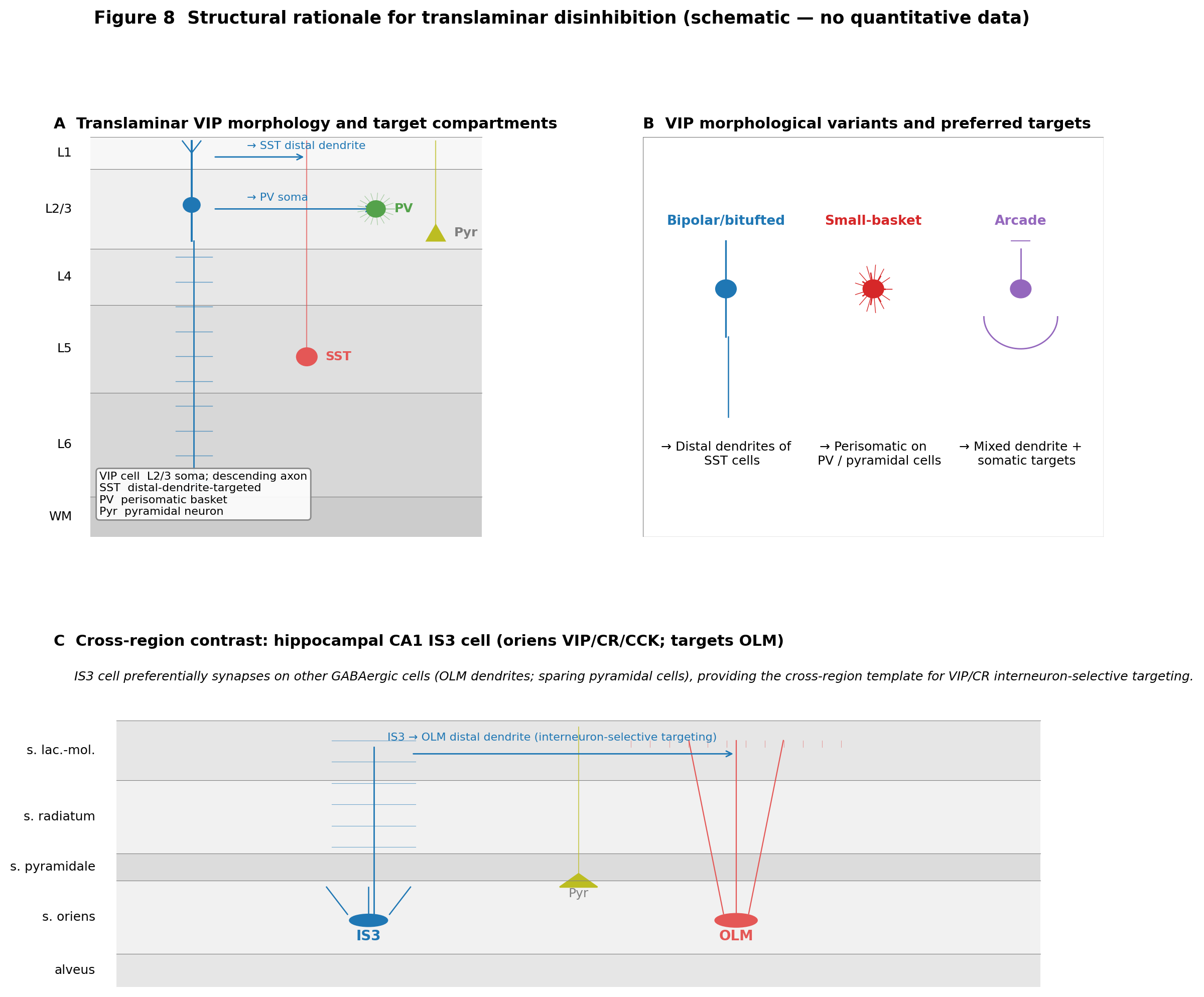
Figure 6:Structural rationale for translaminar disinhibition (schematic). (A) Idealised L2/3-soma + descending-axon VIP cell drawn over a six-layer cortical column, with SST, PV, and pyramidal postsynaptic compartments highlighted; based on the canonical morphological repertoire described by Kawaguchi & Kubota (1996), Prönneke et al. (2015), and Apicella & Marchionni (2022) and the connectomic/Patch-seq target distributions reported by Lee et al. (2013), Zhou et al. (2017), Schneider-Mizell et al. (2025), and Gamlin et al. (2025). (B) Comparison cartoon of small-basket, arcade, and bipolar VIP variants, with the postsynaptic compartments each preferentially contacts (perisomatic for small-basket; mixed dendritic/somatic for arcade; distal-dendrite for bipolar/bitufted). (C) Hippocampal IS3 cell schematic (soma in stratum oriens; axon targeting OLM cells) for cross-region contrast. Note: This figure is a schematic with no quantitative data substrate; cited works support the qualitative compartmental relationships only and not any numerical claims.
📓 Figure code
import matplotlib.pyplot as plt
from matplotlib.patches import Rectangle, Circle, Polygon
import numpy as np

fig = plt.figure(figsize=(13.5, 11))
gs = fig.add_gridspec(2, 2, hspace=0.32, wspace=0.28, height_ratios=[1.2, 1.0])

# Panel A: column with VIP cell + targets
axA = fig.add_subplot(gs[0, 0]); axA.set_xlim(0,1); axA.set_ylim(0,1); axA.axis("off")
axA.set_title("A  Translaminar VIP morphology and target compartments",
              loc="left", fontsize=11, fontweight="bold")
for (lo,hi,name), c in zip(
        [(0.92,1.00,"L1"),(0.72,0.92,"L2/3"),(0.58,0.72,"L4"),(0.36,0.58,"L5"),(0.10,0.36,"L6"),(0.00,0.10,"WM")],
        ["#f7f7f7","#efefef","#e7e7e7","#dfdfdf","#d7d7d7","#cccccc"]):
    axA.add_patch(Rectangle((0.08,lo), 0.85, hi-lo, color=c, lw=0))
    axA.text(0.04, (lo+hi)/2, name, ha="right", va="center", fontsize=9)
axA.add_patch(Circle((0.30, 0.83), 0.018, color="#1f77b4"))
axA.plot([0.30, 0.30], [0.85, 0.99], color="#1f77b4", lw=1.4)
axA.plot([0.30, 0.30], [0.81, 0.74], color="#1f77b4", lw=1.4)
axA.plot([0.305, 0.305], [0.74, 0.12], color="#1f77b4", lw=1.0)
axA.add_patch(Circle((0.55, 0.45), 0.022, color="#e45756")); axA.text(0.59, 0.45, "SST", color="#e45756", fontweight="bold")
axA.add_patch(Circle((0.70, 0.82), 0.020, color="#54a24b")); axA.text(0.74, 0.82, "PV", color="#54a24b", fontweight="bold")
axA.add_patch(Polygon([(0.83, 0.78), (0.81, 0.74), (0.85, 0.74)], color="#bcbd22"))

# Panel B: variants
axB = fig.add_subplot(gs[0, 1]); axB.set_xlim(0,1); axB.set_ylim(0,1); axB.axis("off")
axB.set_title("B  VIP morphological variants and preferred targets",
              loc="left", fontsize=11, fontweight="bold")
axB.text(0.18, 0.5, "Bipolar/bitufted\n-> SST distal", ha="center", fontsize=10, color="#1f77b4")
axB.text(0.50, 0.5, "Small-basket\n-> PV/Pyr soma", ha="center", fontsize=10, color="#d62728")
axB.text(0.82, 0.5, "Arcade\n-> mixed", ha="center", fontsize=10, color="#9467bd")

# Panel C: hippocampal IS3 -> OLM
axC = fig.add_subplot(gs[1, :]); axC.set_xlim(0,1); axC.set_ylim(0,1); axC.axis("off")
axC.set_title("C  Cross-region: hippocampal CA1 IS3 -> OLM (interneuron-selective)",
              loc="left", fontsize=11, fontweight="bold")
for (lo,hi,name), c in zip(
        [(0.0,0.10,"alveus"),(0.10,0.32,"oriens"),(0.32,0.40,"pyramidale"),(0.40,0.62,"radiatum"),(0.62,0.80,"lac.-mol.")],
        ["#e6e6e6","#f1f1f1","#dcdcdc","#f1f1f1","#e6e6e6"]):
    axC.add_patch(Rectangle((0.06,lo), 0.88, hi-lo, color=c, lw=0))
    axC.text(0.04, (lo+hi)/2, name, ha="right", va="center", fontsize=9)
axC.add_patch(Circle((0.30, 0.20), 0.018, color="#1f77b4"))
axC.add_patch(Circle((0.65, 0.20), 0.020, color="#e45756"))

fig.suptitle("Figure 8  Structural rationale for translaminar disinhibition (schematic)",
             fontsize=12.5, y=0.995, fontweight="bold")
fig.savefig("fig-vip-axonal-targeting-schematic.png", dpi=200, bbox_inches="tight")
fig.savefig("fig-vip-axonal-targeting-schematic.pdf", bbox_inches="tight")
plt.close(fig)
Several methodological caveats apply to within-VIP morphological partitions. First, all of the major rodent reconstructed datasets — a foundational study, Gouwens et al. (2019), Gouwens et al. (2020), an earlier report, and a preceding study — rely on VIP-Cre genetic labelling, which captures the Vip-expressing transcriptomic subclass but does not perfectly resolve VIP-peptide expression on a cell-by-cell basis; the difference matters most for rodent-to-primate translation, where the calretinin-positive analogue is the operational handle Medalla et al., 2023Hodge et al., 2019Fishell & Kepecs, 2020. Second, axonal completeness varies substantially across the field: in vitro slice biocytin reconstructions truncate distal axon collaterals, while EM reconstructions are constrained by the volume — together producing artefactual differences in laminar axon distributions that confound met-type definition Gouwens et al., 2020Schneider-Mizell et al., 2025Elabbady et al., 2025Gliko et al., 2024. Third, the dendrite-emerging-axon feature reported by an initial investigation is sensitive to the resolution of the axon initial segment in the imaging modality, which differs across the studies that have searched for it Badrinarayanan et al., 2021. These are not arguments against the convergent picture; they are constraints on how strongly the discrete-versus-continuum and cross-area conflicts above can be adjudicated from current data.
Summary and forward links
VIP-class morphological diversity is structured but graded. A bipolar/bitufted somatodendritic profile with a vertically oriented, dendrite-emerging, mostly inhibition-targeting axon dominates the supragranular VIP population in rodent neocortex Kawaguchi & Kubota, 1996Prönneke et al., 2015Apicella & Marchionni, 2022Schneider-Mizell et al., 2025. A minority of small-basket, arcade, and multipolar variants — together with the L6 perisomatic-bouton fraction and the deep-layer multipolar repertoire — extends the class beyond the canonical bipolar form Rudy et al., 2010Zhou et al., 2017Apicella & Marchionni, 2022Yayon et al., 2022. Patch-seq and connectomic atlases recover discrete morpho-electric centroids embedded in continuous variation rather than fully discrete classes Gouwens et al., 2020Gouwens et al., 2020Gliko et al., 2024Schneider-Mizell et al., 2025. Cross-area and cross-species shifts (mPFC, MEC, primate V1, human MTG and L1) repeat the overall layout while quantitatively reweighting subtype proportions, dendritic geometries, and biophysical parameters Moreno et al., 2026Badrinarayanan et al., 2021Medalla et al., 2023Chartrand et al., 2023. These morphologies are realised electrically as the heterogeneous intrinsic-firing repertoire surveyed in Section Intrinsic Electrophysiology, and they constrain the synaptic and circuit motifs reviewed in Section Synaptic Properties and Connectivity and revisited in Section the concluding synthesis.
- Apicella, A. junior, & Marchionni, I. (2022). VIP-Expressing GABAergic Neurons: Disinhibitory vs. Inhibitory Motif and Its Role in Communication Across Neocortical Areas. Frontiers in Cellular Neuroscience, 16. 10.3389/fncel.2022.811484
- Rudy, B., Fishell, G., Lee, S., & Hjerling-Leffler, J. (2010). Three groups of interneurons account for nearly 100% of neocortical GABAergic neurons. Developmental Neurobiology, 71(1), 45–61. 10.1002/dneu.20853
- Yayon, N., Amsalem, O., Zorbaz, T., Yakov, O., Dubnov, S., Winek, K., Dudai, A., Adam, G., Schmidtner, A. K., Tessier-Lavigne, M., Renier, N., Habib, N., Segev, I., London, M., & Soreq, H. (2022). High-throughput morphometric and transcriptomic profiling uncovers composition of naïve and sensory-deprived cortical cholinergic VIP/CHAT neurons. The EMBO Journal, 42(1). 10.15252/embj.2021110565
- Gouwens, N. W., Sorensen, S. A., Berg, J., Lee, C., Jarsky, T., Ting, J., Sunkin, S. M., Feng, D., Anastassiou, C. A., Barkan, E., Bickley, K., Blesie, N., Braun, T., Brouner, K., Budzillo, A., Caldejon, S., Casper, T., Castelli, D., Chong, P., … Koch, C. (2019). Classification of electrophysiological and morphological neuron types in the mouse visual cortex. Nature Neuroscience, 22(7), 1182–1195. 10.1038/s41593-019-0417-0
- Gouwens, N. W., Sorensen, S. A., Baftizadeh, F., Budzillo, A., Lee, B. R., Jarsky, T., Alfiler, L., Baker, K., Barkan, E., Berry, K., Bertagnolli, D., Bickley, K., Bomben, J., Braun, T., Brouner, K., Casper, T., Crichton, K., Daigle, T. L., Dalley, R., … Zeng, H. (2020). Integrated Morphoelectric and Transcriptomic Classification of Cortical GABAergic Cells. Cell, 183(4), 935-953.e19. 10.1016/j.cell.2020.09.057
- Gliko, O., Mallory, M., Dalley, R., Gala, R., Gornet, J., Zeng, H., Sorensen, S. A., & Sümbül, U. (2024). High-throughput analysis of dendrite and axonal arbors reveals transcriptomic correlates of neuroanatomy. Nature Communications, 15(1). 10.1038/s41467-024-50728-9
- Elabbady, L., Seshamani, S., Mu, S., Mahalingam, G., Schneider-Mizell, C. M., Bodor, A. L., Bae, J. A., Brittain, D., Buchanan, J., Bumbarger, D. J., Castro, M. A., Dorkenwald, S., Halageri, A., Jia, Z., Jordan, C., Kapner, D., Kemnitz, N., Kinn, S., Lee, K., … Collman, F. (2025). Perisomatic ultrastructure efficiently classifies cells in mouse cortex. Nature, 640(8058), 478–486. 10.1038/s41586-024-07765-7
- Gouwens, N. W., Sorensen, S. A., Baftizadeh, F., Budzillo, A., Lee, B. R., Jarsky, T., Alfiler, L., Arkhipov, A., Baker, K., Barkan, E., Berry, K., Bertagnolli, D., Bickley, K., Bomben, J., Braun, T., Brouner, K., Casper, T., Crichton, K., Daigle, T. L., … Zeng, H. (2020). Toward an integrated classification of neuronal cell types: morphoelectric and transcriptomic characterization of individual GABAergic cortical neurons. openRxiv. 10.1101/2020.02.03.932244
- Prönneke, A., Scheuer, B., Wagener, R. J., Möck, M., Witte, M., & Staiger, J. F. (2015). Characterizing VIP Neurons in the Barrel Cortex of VIPcre/tdTomato Mice Reveals Layer-Specific Differences. Cerebral Cortex, 25(12), 4854–4868. 10.1093/cercor/bhv202
- Chartrand, T., Dalley, R., Close, J., Goriounova, N. A., Lee, B. R., Mann, R., Miller, J. A., Molnar, G., Mukora, A., Alfiler, L., Baker, K., Bakken, T. E., Berg, J., Bertagnolli, D., Braun, T., Brouner, K., Casper, T., Csajbok, E. A., Dee, N., … Lein, E. S. (2023). Morphoelectric and transcriptomic divergence of the layer 1 interneuron repertoire in human versus mouse neocortex. Science, 382(6667). 10.1126/science.adf0805
- Kawaguchi, Y., & Kubota, Y. (1996). Physiological and morphological identification of somatostatin- or vasoactive intestinal polypeptide-containing cells among GABAergic cell subtypes in rat frontal cortex. The Journal of Neuroscience, 16(8), 2701–2715. 10.1523/jneurosci.16-08-02701.1996
- Cauli, B., Audinat, E., Lambolez, B., Angulo, M. C., Ropert, N., Tsuzuki, K., Hestrin, S., & Rossier, J. (1997). Molecular and Physiological Diversity of Cortical Nonpyramidal Cells. The Journal of Neuroscience, 17(10), 3894–3906. 10.1523/jneurosci.17-10-03894.1997
- Cauli, B., Zhou, X., Tricoire, L., Toussay, X., & Staiger, J. F. (2014). Revisiting enigmatic cortical calretinin-expressing interneurons. Frontiers in Neuroanatomy, 8. 10.3389/fnana.2014.00052
- Vucurovic, K., Gallopin, T., Ferezou, I., Rancillac, A., Chameau, P., van Hooft, J. A., Geoffroy, H., Monyer, H., Rossier, J., & Vitalis, T. (2010). Serotonin 3A Receptor Subtype as an Early and Protracted Marker of Cortical Interneuron Subpopulations. Cerebral Cortex, 20(10), 2333–2347. 10.1093/cercor/bhp310
- Stachniak, T. J., Kastli, R., Hanley, O., Argunsah, A. Ö., van der Valk, E. G. T., Kanatouris, G., & Karayannis, T. (2021). Postmitotic Prox1 Expression Controls the Final Specification of Cortical VIP Interneuron Subtypes. The Journal of Neuroscience, 41(39), 8150–8162. 10.1523/jneurosci.1021-21.2021