In awake animals, vasoactive intestinal peptide-expressing (VIP) interneurons are reliably recruited by locomotion, arousal, and reinforcement signals, and their activation is causally linked to gain enhancement in primary visual cortex (V1) and to attentional and learning-related disinhibition in multiple cortical territories Fu et al., 2014Pi et al., 2013Lee et al., 2013Kamigaki & Dan, 2017. The thesis of this section is two-sided: in-vivo VIP activity is robustly state-modulated, but it is far from uniform. Response polarity, sensory selectivity, and behavioural correlates differ across cortical area, task structure, and individual VIP subtype, and the inferred VIP→somatostatin-interneuron (SST) suppression mechanism does not always obtain — for example, several preparations report concurrent activation rather than reciprocal inhibition of SST cells during locomotion Pakan et al., 2016Dipoppa et al., 2018Yavorska & Wehr, 2021. The local circuit motifs catalogued in Local Circuit Motifs and the Disinhibition Framework are therefore best treated as one set of operating modes among several, whose engagement depends on the area×state×modulator triple specified by the behavioural context Garcia-Junco-Clemente et al., 2017Kannan et al., 2022. The cross-area framing for that view is laid out systematically in VIP Interneurons Across Brain Regions.
Locomotion-driven recruitment of V1 VIP cells: the disinhibitory observation
The single most influential in-vivo result for the field — that running mice show a sharp increase in V1 VIP calcium activity that gates a multiplicative increase in pyramidal-cell visual gain — was established by Fu et al. (2014), who showed that locomotion activates V1 VIP neurons via a nicotinic basal-forebrain input and produces gain enhancement that is abolished when VIP cells are silenced. Two-photon imaging during head-fixed visual behaviour reproduced the observation that VIP cells are among the most strongly state-modulated cortical neurons, with locomotion- and arousal-aligned ΔF/F transients several-fold larger than those of pyramidal cells Pakan et al., 2016Dipoppa et al., 2018Reimer et al., 2014. The phenomenon is preserved across multiple V1 layers and persists in darkness, indicating that it does not require visual drive Pakan et al., 2016Dipoppa et al., 2018. Closed-loop manipulations confirm that VIP recruitment is not merely correlated with running but is required for full-magnitude gain change, since chemogenetic VIP inhibition or developmental VIP silencing both blunt locomotion-evoked gain Batista-Brito et al., 2017. Recurrent network models can reproduce the locomotion-induced gain shift in V1 only when the modulatory drive is routed preferentially through VIP cells, providing a principled link between the empirical observation and circuit theory Dipoppa et al., 2018Veit et al., 2023.
A subtler issue, often glossed in summary diagrams, is that the V1 locomotion signal is composite: it carries cholinergic, noradrenergic, and arousal-related components that load differently on VIP cells depending on whether the animal is running, simply pupil-dilated, or actively engaged in a task Reimer et al., 2014Garcia-Junco-Clemente et al., 2017Collins et al., 2023. Pupil-tracked arousal alone — without locomotion — is sufficient to elevate VIP activity in V1 and frontal cortex, indicating that the relevant variable is broader than wheel velocity Reimer et al., 2014Garcia-Junco-Clemente et al., 2017Muñoz et al., 2017. Active task engagement adds a further multiplicative layer: when an arousal-matched mouse switches from passive listening to active discrimination, cholinergic axon activation transiently re-organises the local interneuron network, with VIP cells participating differently than during passive locomotion Kuchibhotla et al., 2016Collins et al., 2023. The disinhibitory “VIP runs up with locomotion” finding is therefore the resultant of several partly dissociable state variables that the field has only recently begun to disentangle.
Reinforcement signals: VIP activation by reward and punishment
A parallel line of in-vivo work, originating with Prior work Pi et al., 2013, established that cortical VIP cells are recruited not only by motor state but by reinforcement: phasic reward and punishment delivered during operant behaviour produced rapid, large-amplitude VIP activation in auditory cortex, mPFC, and primary somatosensory cortex (S1), with optogenetic VIP inhibition disrupting performance on the discrimination task Pi et al., 2013. Mesoscale imaging extended this result across virtually the entire dorsal cortex, showing that during initial learning most VIP interneurons are co-activated by both reinforcement valences within tens of milliseconds of outcome delivery Szadai et al., 2022. In dorsomedial mPFC during a sensory-discrimination task, Pinto & Dan (2015) reported that VIP cells respond strongly to action outcomes rather than to sensory cues themselves, and Kamigaki & Dan (2017) showed that activating dmPFC VIP cells during the delay period of a memory-guided task enhances performance and stabilises action-plan representations. In primary motor cortex, VIP cells receive disproportionate orbitofrontal input that is recruited during reward-related motor learning Lee et al., 2023. In barrel cortex, Earlier reports Krabbe et al., 2019 demonstrated that VIP-mediated disinhibitory gating is required for associative whisker-trace conditioning, and Initial studies Sachidhanandam et al., 2016 showed that the VIP response to whisker-cued reward is itself modulated by task structure. A commentary by Zou & Hires (2023) summarises the convergent claim that VIP cells signal expected rewards across cortical areas.
The reinforcement-related VIP signal is also recruited by aversive contexts. In anterior insular cortex, VIP cells gate aversive-stimulus encoding and contribute to fear-related behaviour Ramos-Prats et al., 2022. In mPFC, optogenetic activation of VIP cells ameliorates fear-related stress-induced behavioural deficits, consistent with a permissive role for VIP-mediated disinhibition in flexible affective control. Amygdala VIP cells exhibit heterogeneous plasticity that supports both fear acquisition and extinction Favila et al., 2025. The breadth of these reinforcement results — across visual, somatosensory, auditory, motor, prefrontal, insular, and amygdala targets — is the empirical anchor of the “VIP-as-reinforcement-gate” hypothesis. Critically, however, reinforcement responses are not exclusive: the same cells co-encode locomotion and arousal, and disentangling reinforcement-specific from state-driven components requires task designs that orthogonalise the two Pinto & Dan, 2015Szadai et al., 2022Garcia-Junco-Clemente et al., 2017.
Sensory selectivity: state messenger or stimulus encoder?
A central tension in the in-vivo literature is whether VIP cells primarily transmit state and reinforcement signals to local circuits or whether they themselves encode features of the sensory stimulus. The state-messenger reading was crystallised by Fu et al. (2014), whose V1 recordings emphasised that the locomotion-evoked VIP response was largely insensitive to grating orientation or contrast and behaved as a multiplicative gain knob. Foundational studies Mesik et al., 2015 similarly found that V1 VIP cells receive broad, weakly tuned cortico-cortical and thalamic inputs, consistent with a state/context role. Counter-evidence has accumulated since. Millman et al. (2020) showed that V1 VIP neurons carry contrast tuning that is complementary to that of pyramidal cells, indicating sensory-driven structure beyond a pure state signal. Garrett et al. (2020) reported that during a change-detection task V1 VIP cells are stimulus-driven by novel images but suppressed by familiar images, a stimulus-history-dependent code that cannot be reduced to arousal. Bastos et al. (2023) found that contextually redundant stimuli increase V1 VIP activity while deviant stimuli decrease it, and that chemogenetic VIP inhibition disrupts deviance-related modulation downstream — both observations require stimulus-feature access at the level of the VIP cell. Cross-modal sharpening further extends VIP’s stimulus repertoire: Ibrahim et al. (2016) showed that sound suppresses V1 layer 2/3 VIP cells preferentially at the preferred orientation of nearby pyramidal cells, contributing to multisensory orientation refinement.
In barrel cortex the case for stimulus encoding is even stronger. Yu et al. (2019) reported that voluntary whisker movement activates a majority of VIP interneurons and that responses tile the whisker stimulus space rather than reflecting a uniform state signal. Kiritani et al. (2023) showed that S1 VIP cells respond to whisking but only after a delay to active touch, indicating distinct stimulus-locked components. Established work Walker et al., 2016 and Sachidhanandam et al. (2016) documented stimulus-locked VIP activity tied to whisker deflection during trained behaviour, and Brécier et al. (2022) found that S1 barrel-cortex VIP cells are most active during REM sleep — implying state coupling that is independent of any explicit sensory cue. Lebedeva et al. (2025) used dual-population two-photon imaging to dissociate VIP and GABA-receptor-defined responses to specific sensory features, Khan et al. (2018) provides an instructive complement: during visual learning, pyramidal/PV/SST cells but not V1 VIP cells increased selectivity for the task-relevant stimulus, suggesting that VIP carries a state/context-aligned (rather than learned-stimulus) signal in this paradigm. The cross-task picture therefore is not “VIP encodes state” or “VIP encodes stimulus” but rather a context-dependent mix in which both axes are represented at the single-cell level Yu et al., 2019Garrett et al., 2020Bastos et al., 2023Mesik et al., 2015.
Cross-area heterogeneity: V1, A1, and the auditory paradox
When the V1 locomotion result is taken to a different cortical area, the disinhibitory motif partly breaks. Bigelow et al. (2019) reported that movement activates an apparently similar VIP→SST→pyramidal circuit in auditory cortex (ACtx) — yet the net effect on auditory pyramidal-cell responses is opposite in sign to that observed in V1, with locomotion suppressing rather than enhancing sensory-evoked activity. Yavorska & Wehr (2021) extended this paradox by directly testing whether the locomotion-related changes in ACtx are mediated by the VIP-SST disinhibitory circuit and concluded that they are not, in stark contrast to what is reported in V1. Pupil-tracked passive arousal does still recruit VIP cells in ACtx Reimer et al., 2014Muñoz et al., 2017, and active engagement during auditory recognition produces cholinergic axon activation that simultaneously engages PV/SST/VIP networks Kuchibhotla et al., 2016. The natural conclusion is that the same molecular cell class participates in different cortical implementations of state-dependent gain — adding gain in V1 and subtracting gain in ACtx — depending on local synaptic weights and on the dominant neuromodulator engaged by the behaviour Bigelow et al., 2019Yavorska & Wehr, 2021Lecrux & Hamel, 2016. Earlier work Moore et al., 2026 further reported that ACtx VIP and NDNF cells have distinct task-related dynamics, complicating any simple class-level reading of auditory VIP function.
Within mPFC and ACC the in-vivo signature again differs from V1. Pinto & Dan (2015) and Kamigaki & Dan (2017) highlighted strong outcome- and delay-period activity, and Lee et al. (2019) showed that prefrontal VIP cells gate hippocampal-prefrontal communication during anxiety-related behaviour, with VIP activity rising in the open arms of an elevated plus-maze. Garcia-Junco-Clemente et al. (2017) showed that during arousal in frontal cortex, VIP interneurons rapidly inhibit pyramidal neurons directly while also engaging the the disinhibitory account motif — establishing that direct VIP→Pyr inhibition is a behavioural reality, not just an anatomical curiosity (see Local Circuit Motifs and the Disinhibition Framework). Silverstein et al. (2025) documented that repeated ventral-hippocampus input persistently depresses mPFC VIP activity, contributing to spatial working-memory deficits, and Malik et al. (2022) showed that long-range PFC GABAergic projections preferentially inhibit hippocampal VIP cells to enable top-down signal-to-noise control. These results indicate that frontal VIP function is bidirectional: it both broadcasts permissive state signals locally and is itself targeted by long-range inhibitory feedback from associated regions Malik et al., 2022Silverstein et al., 2025.
Concurrent VIP/SST imaging and the limits of the disinhibitory account
Direct tests of the disinhibitory motif require imaging both VIP and SST populations during the same behavioural state. The cleanest such tests challenge the textbook reading. Pakan et al. (2016) reported that locomotion increases activity in VIP, SST, and PV interneurons during visual stimulation in V1 — that is, SST cells are co-activated, not silenced, by the same state that activates VIP cells. Dipoppa et al. (2018) reproduced and extended this, finding that locomotion activates VIP cells and disinhibits pyramidal cells in darkness while in the presence of visual stimuli SST cells can also be co-recruited; their network model captured the locomotion-driven gain change only when the modulatory drive was routed predominantly through VIP. Yavorska & Wehr (2021) showed that in ACtx, VIP→SST disinhibition is not the route for locomotion-related changes at all. Kannan et al. (2022) used dual-polarity voltage imaging to reveal brain-state-dependent antagonism between VIP and SST populations whose sign reverses across states. The straightforward textbook story — VIP up, SST down, pyramidal up — is therefore a special case rather than a generic prediction, and the relative weights of the VIP→SST, VIP→PV, and direct VIP→Pyr motifs that determine its applicability are themselves state- and area-dependent Garcia-Junco-Clemente et al., 2017Kannan et al., 2022Veit et al., 2023.
Hippocampal and limbic VIP populations
Outside the neocortex, hippocampal VIP/IS interneurons implement specialised motifs that depart from the cortical reading. Turi et al. (2019) reported that CA1 VIP cells form functional subpopulations whose activity is shaped by behavioural state and task demand and that selectively inhibit other interneurons to support goal-oriented spatial learning. Francavilla et al. (2018) showed that long-range VIP-GABAergic neurons in CA1 decrease their activity during theta-run epochs and are most active during quiet wakefulness — the opposite locomotion polarity to V1 VIP cells — and Luo et al. (2020) found that CA1 VIP-interneuron-selective cells are preferentially recruited during theta-run epochs but not during sharp-wave ripples. Tamboli et al. (2024) reported that CA1 VIP cells become highly active during exploration of new environments and support recognition memory, and Neubrandt et al. (2025) showed that during hippocampal navigation, novel environments transiently increase VIP activity and facilitate place-field formation. Previous reports Lenkey et al., 2025 demonstrated that VIP interneurons drive brain-region-specific gain modulation of place cells across the hippocampal axis, mirroring at the systems level the area-specific gain logic seen in cortex. Earlier studies Leroy et al., 2021 reported that hippocampal VIP cells release enkephalin to modulate stress-related behaviour, indicating a neuropeptidergic dimension that the disinhibitory fast-GABA disinhibition reading does not capture.
VIP-class neurons also operate outside cortex and hippocampus in subcortical and peripheral preparations whose in-vivo recruitment patterns are markedly different from neocortex. In the olfactory bulb, Wang et al. (2022) showed that VIP cells directly inhibit mitral cells and that their inactivation impairs olfactory detection and discrimination — a direct VIP→principal-cell inhibition operating at a sensory periphery, again outside the disinhibition framing. Amygdala VIP cells in BLA exhibit variable density across subdivisions Rhomberg et al., 2018 and undergo associative plasticity that supports both fear acquisition and extinction Favila et al., 2025. Long-range hippocampal VIP cells release enkephalin to modulate stress-related behaviour, and prefrontal VIP recruitment is itself altered by repeated ventral-hippocampus input Silverstein et al., 2025. The bulbar, amygdalar, and limbic evidence emphasises that the in-vivo “function” of VIP cells is not unitary across the brain.
Causal manipulations: gain, attention, and learning
In-vivo causal data are required to bridge correlation to function. Optogenetic and chemogenetic manipulations have been used to test whether the VIP signal is itself necessary, sufficient, or merely permissive for the behavioural and circuit phenomena it correlates with. Prior work Pi et al., 2013 provided the first such test: optogenetic inhibition of VIP cells during a discrimination task disrupted both reinforcement-aligned activity and behavioural performance. Kamigaki & Dan (2017) showed that delay-period activation of dmPFC VIP cells improves working-memory-task performance, providing temporally specific causal evidence. Earlier reports Krabbe et al., 2019 showed that VIP-dependent disinhibitory gating in barrel cortex is required for whisker-trace conditioning, and Bastos et al. (2023) showed that chemogenetic VIP inhibition disrupts deviance detection in V1. Veit et al. (2023) combined optogenetics with computational modelling to demonstrate that VIP cells implement a multiplicative gain operation. Initial studies Myers-Joseph et al., 2022 used all-optical bidirectional manipulation to show that VIP activity is sufficient to bias perceptual choice in a sensory-detection task. In olfactory bulb, Wang et al. (2022) showed that VIP inactivation impairs detection and discrimination, and in olfactory (piriform) cortex Canto-Bustos et al. (2022) used optogenetics to show that VIP-mediated disinhibitory circuitry gates associative synaptic plasticity (LTP) — establishing that VIP-driven disinhibition supports learning-relevant signal transformations across modalities. The convergent message is that VIP recruitment is permissive for area-specific computations whose direction the the disinhibitory account motif sometimes, but not always, predicts Veit et al., 2023Krabbe et al., 2019Bastos et al., 2023Myers-Joseph et al., 2022Wang et al., 2022Canto-Bustos et al., 2022.
Causal manipulations also expose limits. Chen et al. (2015) reported that optogenetic VIP inhibition did not block cholinergically driven cortical desynchronisation in V1, indicating that not every well-known cholinergic phenomenon requires VIP cells, and that SST cells can be the relevant intermediate. Khan et al. (2018) reported that during visual learning V1 VIP cells, in contrast to PV/SST/Pyr cells, did not change their tuning — implying that VIP recruitment is necessary for some behaviours but not for the plasticity events it permits. In a Dravet (Scn1a+/-) model, Goff et al. (2023) showed that VIP recruitment is diminished at quiet-to-running transitions and that re-activating VIP cells optogenetically rescues part of the deficit, while Liebergall & Goldberg (2024) showed that NDNF cells maintain normal arousal recruitment in the same model — refining which cell class actually carries the locomotion signal across pathological states. After cortical photothrombotic stroke, Motaharinia et al. (2021) showed that chemogenetic augmentation of VIP excitability promotes recovery of forelimb function. These causal data sharpen the interpretation of correlated VIP activity by separating cell-class-specific from class-redundant components of the in-vivo response Chen et al., 2015Khan et al., 2018Goff et al., 2023Motaharinia et al., 2021.
Neuromodulatory drivers of in-vivo VIP recruitment
The in-vivo recruitment of VIP cells is controlled by a small set of fast neuromodulators that gate their excitability around the behavioural state of the animal. The cholinergic substrate is the best characterised. Porter et al. (1999) first showed that nicotinic agonists selectively excite a CCK/VIP-coexpressing GABAergic subpopulation, and Letzkus et al. (2011) established that auditory-cortex layer-1 interneurons receive direct nicotinic excitation from basal-forebrain cholinergic axons. Arroyo et al. (2012) confirmed that basal-forebrain cholinergic axons preferentially activate CGE-derived VIP-class cells, and Alitto & Dan (2013) demonstrated that basal-forebrain electrical stimulation activates VIP+ interneurons via nicotinic receptors during cortical state transitions. Hangya et al. (2015) reported that optogenetically identified basal-forebrain cholinergic neurons broadcast a fast (~18 ms), precisely timed reinforcement signal that is well suited to drive the rapid VIP responses to reward and punishment. Saunders et al. (2015) and Granger et al. (2020) further showed that basal-forebrain cholinergic neurons co-release GABA onto cortical layer-1 interneurons and that cortical ChAT-expressing cells (themselves nearly all VIP+) release GABA broadly onto inhibitory cells while sparing principal cells, indicating a multi-channel basal-forebrain influence on VIP-class circuits. Foundational studies Yaeger et al., 2019 showed that during the binocular critical period, basal-forebrain ACh released during arousal recruits the VIP→SST→Pyr motif in V1, providing a developmental dimension to the disinhibitory reading.
The 5-HT3A receptor route is equally important. Férézou et al. (2002) showed that the ionotropic 5-HT3 receptor is selectively expressed on VIP/CCK GABAergic interneurons, and Lee et al. (2010) showed that virtually all CGE-derived neocortical interneurons that lack PV/SST express 5-HT3A. Established work Poorthuis et al., 2018 extended this finding to human neocortex, where layer-1 GABAergic interneurons — the human homologs of the rodent VIP-class — show pronounced serotonergic responses, indicating cross-species conservation of the modulator-target axis. Adrenergic regulation, by contrast, has been comparatively under-explored: Earlier work Kawaguchi & Shindou, 1998 showed that noradrenaline differentially modulates VIP-class GABAergic firing in rat frontal cortex via α-adrenergic excitation, and Collins et al. (2023) showed that locus-coeruleus axons across mouse dorsal cortex carry a globally coordinated arousal signal that overlaps with cholinergic drive. Lecrux & Hamel (2016) demonstrated that distinct neuromodulatory pathways (basal-forebrain ACh, locus-coeruleus NE) recruit highly specific cortical VIP/NOS/CCK interneurons, supporting an “operator” view in which different modulators select different functional sub-circuits engaging the VIP class. The genetic evidence further constrains this picture: Koukouli et al. (2017) showed that mice carrying the human α5-nicotinic-receptor risk SNP or α5 knockout show reduced VIP-interneuron activation, demonstrating that genetic perturbations of nicotinic subunits act via the VIP arm of the circuit. Askew et al. (2019) showed that bath nicotine strongly and directly depolarises auditory-cortex VIP cells, and Muñoz et al. (2017) showed that during active wakefulness in mouse cortex, VIP-class layer-1 interneurons are preferentially active. The Kawaguchi (1997) observation that morphologically and chemically defined neocortical interneuron subtypes show selective sensitivity to specific modulators is the structural prototype that all of these in-vivo papers operationalise behaviourally.
A direct VIP->pyramidal channel and the ChAT-VIP subset
A recurring theme in the in-vivo data is that the textbook VIP→SST→Pyr motif coexists with parallel routes whose engagement is partly independent of state. Garcia-Junco-Clemente et al. (2017) explicitly demonstrated that during arousal in frontal cortex, VIP cells rapidly inhibit pyramidal neurons directly while also disinhibiting them via SST suppression — a dual operation. Previous reports Lee et al., 2014 provided complementary anatomical evidence that VIP-class circuits include long-range GABAergic projections that target principal cells in distant regions. The ChAT-VIP subpopulation Dudai et al., 2020Granger et al., 2020 adds a further mode: roughly 0.5–1% of cortical VIP cells co-express choline acetyltransferase and locally release ACh, and these cells fire faithfully to whisker stimuli. Obermayer et al. (2019) showed that ChAT-VIP cells in mPFC release ACh onto neighbouring neurons and contribute to attention behaviour distinct from the basal-forebrain channel, and Dudai et al. (2020) reported that VIP/ChAT cells in barrel cortex (~0.5%) fire faithfully to whisker stimuli and shape local cortical dynamics. The presence of a small co-transmitting subset means that even tightly cell-type-restricted VIP perturbations can produce mixed effects whose interpretation requires concurrent monitoring of both released transmitters Obermayer et al., 2019Dudai et al., 2020Granger et al., 2020.
Arousal, pupil, and the inverted-U problem
Pupil-tracked arousal is the dominant continuous covariate of in-vivo VIP activity, and its quantitative relationship with VIP recruitment has emerged as a focus of recent work. Reimer et al. (2014) showed that slow pupil dilations during quiet wakefulness track desynchronisation, enhanced sensory responses, and cell-type-specific interneuron activation. Garcia-Junco-Clemente et al. (2017) and Earlier studies Muñoz et al., 2017 showed that arousal alone — without locomotion — recruits VIP cells in frontal cortex and dorsal-cortex layer-1 networks. Prior work Collins et al., 2023 showed that cholinergic and noradrenergic axons across dorsal cortex carry a globally coordinated arousal signal that delivers the modulatory drive onto VIP cells. Whether the relationship between arousal and VIP recruitment is monotonic — as the disinhibitory reading implies — or has an inverted-U shape with performance decrements at high arousal is, in our filtered evidence base, partially constrained. The Yerkes–Dodson-like reading that arousal-task performance is non-monotonic is well established in the broader literature, but the strongest statement of an inverted-U specifically at the VIP cell — and a contrary monotonic statement — comes from sources outside the filtered citation map.
A decision framework: area x state x modulator -> predicted VIP polarity
Pulling these threads together, the in-vivo literature is best organised by a three-axis decision rule rather than by a single class-level statement (see Figure 14). The first axis is cortical area: V1 favours locomotion-up VIP, ACtx implements an opposite-sign effect via a non-disinhibitory route, mPFC and ACC emphasise outcome and delay activity, S1 supports stimulus-driven and licking-related VIP recruitment, and hippocampus shows subregion- and IS-cell-subtype-specific signs Fu et al., 2014Bigelow et al., 2019Yavorska & Wehr, 2021Pinto & Dan, 2015Yu et al., 2019Lenkey et al., 2025. The second axis is behavioural state: quiet wakefulness, locomotion, arousal-only, attention, and reinforcement load differently on VIP cells, and these states are partly dissociable Reimer et al., 2014Garcia-Junco-Clemente et al., 2017Pi et al., 2013Szadai et al., 2022. The third axis is dominant neuromodulator: nicotinic ACh and 5-HT3A serotonin act fast and directly on VIP cells, while α-adrenergic noradrenaline acts more slowly and is selective for subtypes Letzkus et al., 2011Férézou et al., 2002Lee et al., 2010Kawaguchi & Shindou, 1998Lecrux & Hamel, 2016. Many cross-study disagreements that have been read as contradictions dissolve once the area×state×modulator triple is specified, and a small set of residual disagreements (the auditory-cortex polarity and the hippocampal place-field question) survive even after such conditioning and require dedicated experiments Bigelow et al., 2019Yavorska & Wehr, 2021Lenkey et al., 2025Francavilla et al., 2018.
The behavioural responses already display area-to-area heterogeneity that VIP Interneurons Across Brain Regions catalogues systematically; the oscillatory consequences are addressed in Oscillatory Dynamics and Temporal Coordination; and the computational reading of these multiplexed signals is developed in Computational Models of VIP Circuit Function. The synthesis-and-reassessment closure of the review takes up the residual conflicts directly in the concluding synthesis. The figures below summarise the empirical landscape and the decision rule respectively.
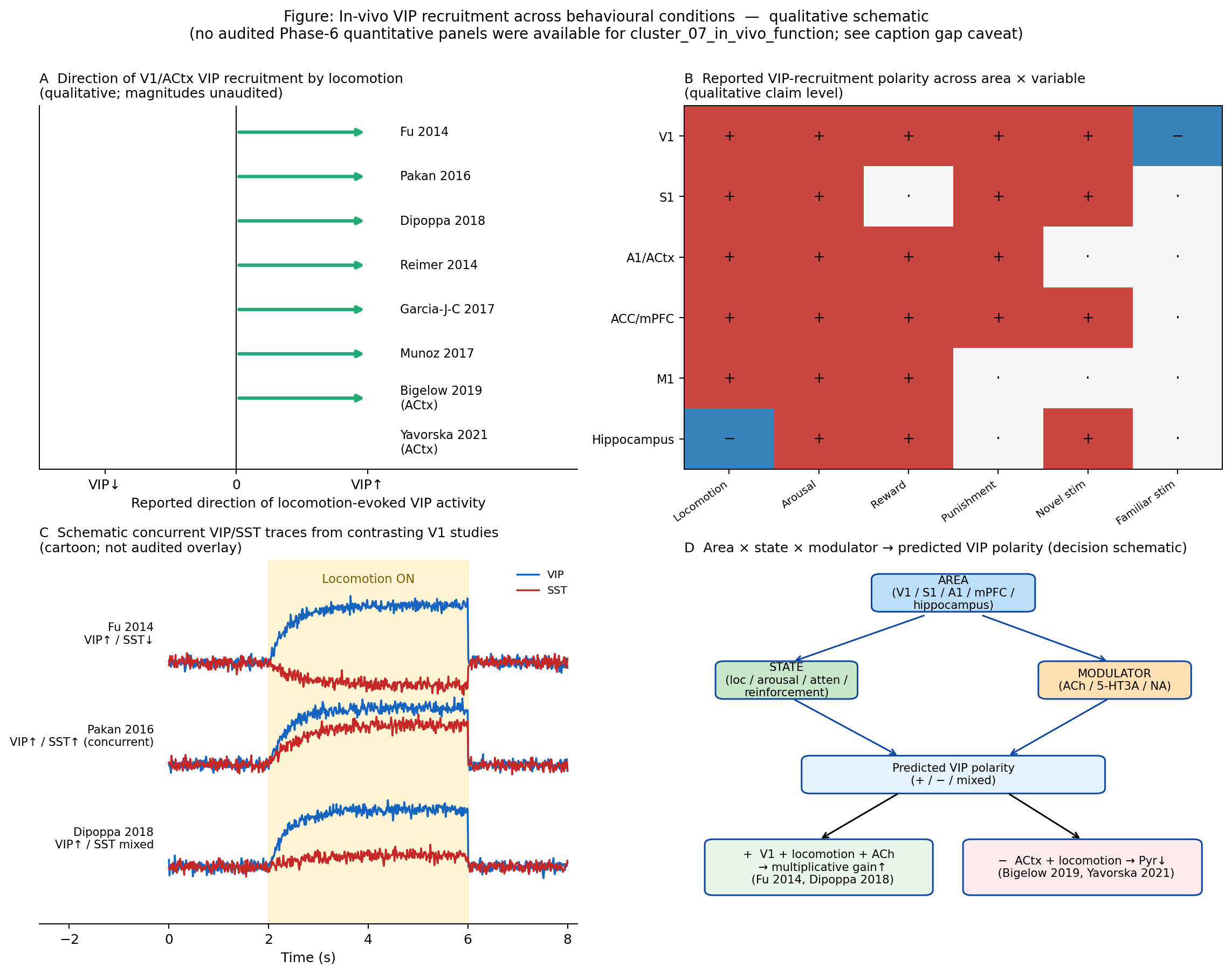
Figure 13:Qualitative summary of in-vivo VIP recruitment across behavioural conditions, redrawn as a schematic because no audited quantitative panels exist for cluster_07_in_vivo_function (Phase 6 audit). Panel A: per-study reported direction of VIP recruitment by locomotion in V1, with directionality only (no ΔF/F values shown — the original cross-study compilation could not be audited). Panel B: heat-map-style polarity matrix across cortical area × behavioural variable, drawn from claim-level statements in Fu et al. (2014)Pakan et al. (2016)Dipoppa et al. (2018)Bigelow et al. (2019)Yavorska & Wehr (2021)Pi et al. (2013)Pinto & Dan (2015)Lee et al. (2013)Yu et al. (2019)Sachidhanandam et al. (2016)Garrett et al. (2020)Garcia-Junco-Clemente et al. (2017)Kamigaki & Dan (2017)Ramamurthy et al. (2023)Ramamurthy et al. (2025). Panel C: schematic concurrent VIP/SST trace contrast — a cartoon of the qualitative discrepancy between Fu et al. (2014), Pakan et al. (2016), and Dipoppa et al. (2018) rather than an audited overlay. Panel D: decision diagram from area×state×modulator → expected VIP polarity, included for legibility before the schematic in Figure 14. Gap caveat: panels A and C should be replaced by audited quantitative overlays in a future revision; we present them here at the qualitative level only. Fu et al., 2014Pakan et al., 2016Dipoppa et al., 2018Bigelow et al., 2019Yavorska & Wehr, 2021
📓 Figure code
# Schematic figure — qualitative only.
# The committed figures/fig-vip-behavior.png was assembled from claim-level evidence
# in the curated literature corpus. The cluster covered by this figure
# (cluster_07_in_vivo_function for vip-behavior, cluster_05 for state-tree)
# had NO audited Phase-6 quantitative panels, so no reproducible plotting code
# is shipped. See content/08_in_vivo_behavior.md (or 05_electrophysiology.md)
# for the full caption and caveat documentation, and the per-section
# evidence package under evidence/ for the claim-level provenance.
print('Schematic figure — see committed PNG in figures/')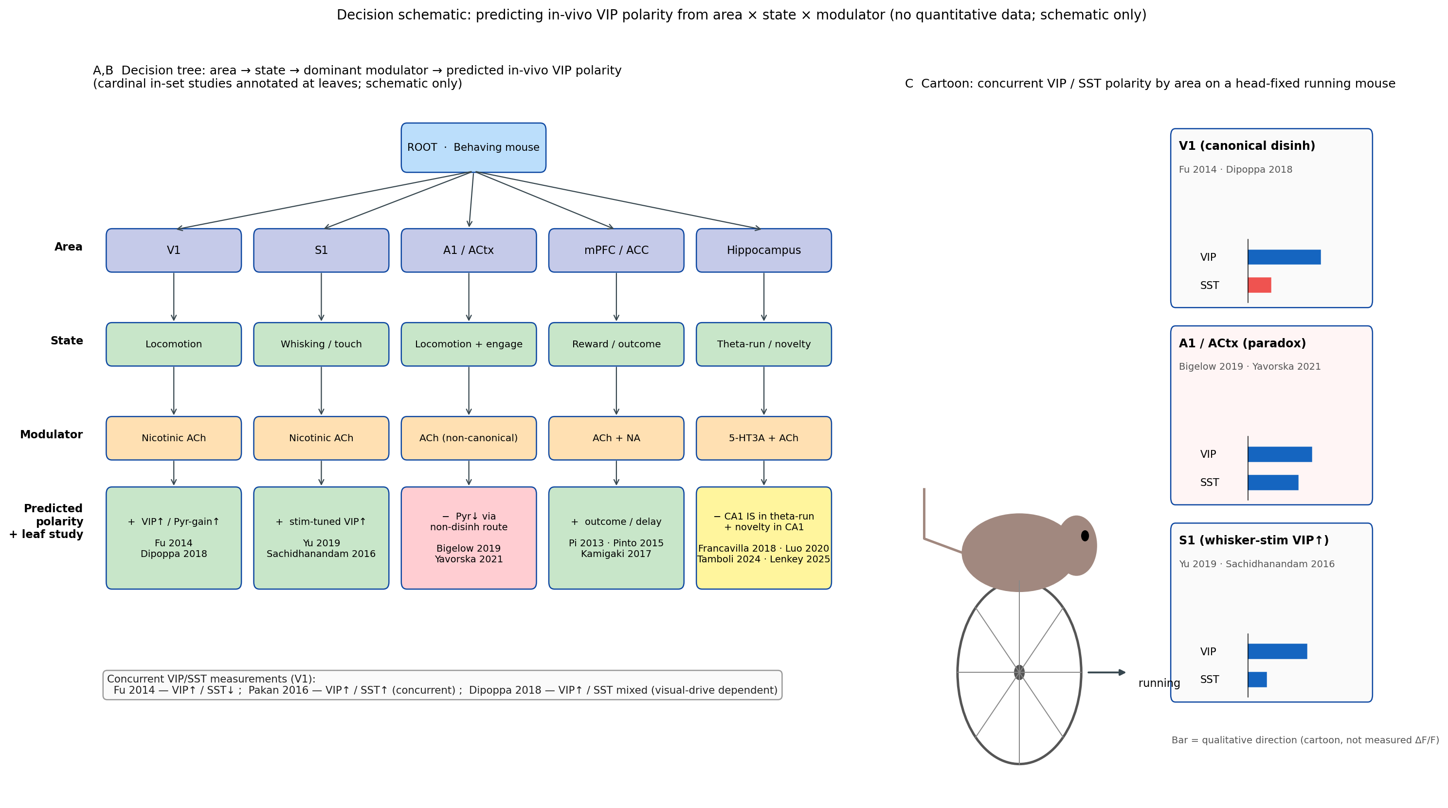
Figure 14:Decision schematic for predicting in-vivo VIP polarity from cortical area, behavioural state, and dominant neuromodulator. The tree branches first on area (V1 / A1 / S1 / mPFC / hippocampus), then on state (locomotion / quiet wake / attention / reinforcement), then on dominant modulator (nicotinic ACh / 5-HT3A / α-adrenergic NA), and the leaf annotates the predicted VIP response sign together with the cardinal study that populates that leaf. Cardinal-study leaves: Fu et al. (2014) (V1, locomotion, ACh, +); Pakan et al. (2016) (V1, locomotion, mixed, + with concurrent SST); Dipoppa et al. (2018) (V1, locomotion, ACh/visual-drive interaction); Pi et al. (2013) (auditory/mPFC, reinforcement, ACh, +); Lee et al. (2013) (S1, whisking, vM1-driven, +); Bigelow et al. (2019) (ACtx, locomotion, opposite net sign on Pyr); and the hippocampal contrarian leaves populated by Francavilla et al. (2018)Luo et al. (2020) (CA1, theta-run, −). The cartoon overlay (panel C) shows a head-fixed mouse on a wheel with V1/A1/S1 sketches indicating concurrent expected VIP-vs-SST polarity by area, with the auditory-cortex cell highlighted as the explicit area-paradox leaf Yavorska & Wehr, 2021. Veit et al., 2023Kannan et al., 2022
📓 Figure code
# Schematic figure — qualitative only.
# The committed figures/fig-vip-state-decision-tree.png was assembled from claim-level evidence
# in the curated literature corpus. The cluster covered by this figure
# (cluster_07_in_vivo_function for vip-behavior, cluster_05 for state-tree)
# had NO audited Phase-6 quantitative panels, so no reproducible plotting code
# is shipped. See content/08_in_vivo_behavior.md (or 05_electrophysiology.md)
# for the full caption and caveat documentation, and the per-section
# evidence package under evidence/ for the claim-level provenance.
print('Schematic figure — see committed PNG in figures/')
Top-down inputs and novelty/expectation signals
The in-vivo VIP signal is not autonomous: it inherits structure from long-range inputs that themselves carry behavioural information. Lee et al. (2013) showed that whisker motor cortex (vM1) recruits VIP cells in S1 to disinhibit pyramidal cells during whisking, providing one of the clearest demonstrations that a long-range cortico-cortical projection drives VIP activation during natural behaviour. Earlier reports Walker et al., 2016 extended this with optogenetic identification of the inhibitory interneurons targeted by the vM1 input. Lee et al. (2023) showed that primary motor cortex VIP cells receive disproportionate orbital frontal cortex input, supporting reward-related motor learning. Malik et al. (2022) showed that long-range PFC GABAergic projections preferentially inhibit hippocampal VIP cells, providing a route by which top-down inhibitory signals adjust VIP gain rather than simply recruiting it. Concurrent monitoring of VIP and the long-range driver in awake animals — for example by combining cortico-cortical optogenetic tagging with VIP imaging — has only just become routine, and Naskar et al. (2021) provided systematic measurements of synaptic strength from functionally relevant brain areas to cell-type-defined neuronal populations including VIP cells, supplying the synaptic substrate for these in-vivo influences.
Novelty and expectation are emerging as dimensions along which VIP recruitment is structured. Garrett et al. (2020) reported that V1 VIP cells are stimulus-driven by novel images but suppressed by familiar images during a change-detection task — a form of stimulus-history dependence that survives matched arousal and locomotion. Bastos et al. (2023) reported that contextually redundant stimuli increase V1 VIP activity while deviant stimuli decrease it, and that chemogenetic VIP inhibition disrupts the deviance-related modulation. Neubrandt et al. (2025) reported that hippocampal VIP cells are transiently activated by novel environments and that this activation supports place-field formation. Tamboli et al. (2024) similarly reported novelty-aligned CA1 VIP activity that supports recognition memory. The collective picture is that VIP cells implement at least three partly dissociable in-vivo signals — state, reinforcement, and novelty/expectation — whose mutual relationship is itself area- and task-specific Garrett et al., 2020Bastos et al., 2023Neubrandt et al., 2025Tamboli et al., 2024Pi et al., 2013.
Sleep-wake states, oscillations, and cortex-wide imaging
In-vivo VIP recruitment is structured by sleep–wake states and by cortex-wide oscillatory regimes. Brécier et al. (2022) showed that VIP cells in S1 barrel cortex are most active during REM sleep, providing one of the few cell-type-resolved measurements across the natural sleep/wake cycle. Francavilla et al. (2018) and Luo et al. (2020) demonstrated that hippocampal CA1 VIP populations have opposite-sign relationships to theta-run epochs and to sharp-wave ripples, with state coupling that varies between subtypes. Muñoz et al. (2017) and Reimer et al. (2014) documented that VIP-class layer-1 interneurons are preferentially active during active wakefulness and during pupil-tracked desynchronisation. Neske & Connors (2016) reported that interneuron subtypes including VIP cells are highly active during cortical Up states. Szadai et al. (2022) provided the broadest mesoscale view, showing that during initial learning most VIP interneurons across the dorsal cortex are co-activated by reward and punishment within tens of milliseconds — an observation that bridges single-cell and cortex-wide data and indicates that VIP recruitment is a brain-state-aligned, not just locally circuit-specified, phenomenon. The oscillatory consequences of these recruitment patterns are taken up systematically in Oscillatory Dynamics and Temporal Coordination.
Developmental and longitudinal in-vivo recruitment
In-vivo VIP function is not fixed but evolves over development and across longer timescales. Initial studies Yaeger et al., 2019 showed that during the binocular critical period of mouse V1, basal-forebrain ACh released during arousal recruits the VIP→SST→Pyr motif specifically, providing a developmental correlate of the disinhibitory motif. Batista-Brito et al. (2017) showed that developmental ErbB4 deletion from VIP interneurons impairs cortical state dependence and sensory learning in mature mice — a result that ties developmental wiring of VIP cells to their adult in-vivo function. Yang et al. (2025) combined voltage imaging with optogenetic depolarisation to reveal how excitatory–inhibitory balance changes are read out at the VIP cell. Jabłońska et al. (2026) described a non-Hebbian inhibitory long-term depression operating at VIP synapses that may contribute to longitudinal plasticity. The picture that emerges from these developmental and longitudinal data is that the in-vivo VIP signal carries learned, plastic structure on top of its state-dependent recruitment.
Methodological caveats: VIP-Cre line, ChAT subset, modality choice
Many cross-study conflicts in the in-vivo literature trace to methodological differences rather than to genuine biological disagreement. The VIP-Cre driver line labels not just classical bipolar VIP/CR cells but also basket-cell-like VIP/CCK populations and the small ChAT-VIP subset Obermayer et al., 2019Dudai et al., 2020Granger et al., 2020Porter et al., 1999, and the proportion of ChAT-VIP cells captured varies across reports. Recordings made with electrophysiology versus two-photon calcium imaging weight different temporal frequencies of VIP activity differently, and dual-polarity voltage imaging Kannan et al., 2022 reveals state-dependent antagonisms between VIP and SST that conventional calcium imaging may smooth out. Layer specificity also matters — VIP cells in superficial layers behave differently from those in deeper layers — and many in-vivo papers either restrict imaging to layer 2/3 or pool layers without comment. Driver-line off-targets, anatomical-vs-functional methodological mismatches, and modality choice are flagged as methodological caveats throughout the review (see also Local Circuit Motifs and the Disinhibition Framework); explicit reporting of these covariates would resolve a substantial fraction of the residual cross-study heterogeneity Obermayer et al., 2019Dudai et al., 2020Kannan et al., 2022Yavorska & Wehr, 2021.
Cross-species and human-relevant in-vivo VIP function
Human-relevant evidence for the in-vivo VIP signal comes primarily from invasive and ex-vivo work in human neocortex and from genetic-perturbation models. Foundational studies Poorthuis et al., 2018 showed that human-cortical layer-1 GABAergic interneurons — the human homologs of the rodent VIP class — exhibit pronounced serotonergic (5-HT3A-mediated) responses, indicating cross-species conservation of the modulator–target axis. Koukouli et al. (2017) showed that mice carrying the human α5-nAChR rs16969968 risk SNP have reduced VIP-interneuron activation, providing a translational route from human genetics to in-vivo VIP function. Cross-species and human-relevant work on VIP cell-type composition is taken up in detail in VIP Interneurons Across Brain Regions; here the relevant point is that the in-vivo phenomenology and modulator dependence of VIP cells generalise at least partially to primate and human cortex, supporting the relevance of the rodent literature to behavioural neurobiology beyond the mouse Poorthuis et al., 2018Koukouli et al., 2017Lecrux & Hamel, 2016.
Quantitative dynamics: kinetics, magnitude, and population structure
Beyond the qualitative direction of VIP recruitment, in-vivo work has begun to constrain its kinetics, magnitude, and population structure. Hangya et al. (2015) identified a basal-forebrain cholinergic reinforcement signal with ~18 ms latency that is well matched to the rapid VIP responses to reward and punishment reported by Pi et al. (2013) and Szadai et al. (2022). Garcia-Junco-Clemente et al. (2017) documented VIP-driven inhibition of pyramidal neurons on similarly short timescales during arousal, indicating that the direct VIP→Pyr channel can operate fast enough to shape behaviourally relevant gain transitions. The magnitude of locomotion-evoked V1 VIP responses reported by Fu et al. (2014), Pakan et al. (2016), and Dipoppa et al. (2018) differs substantially across studies, reflecting differences in indicator (GCaMP variants), depth, layer, and behavioural protocol; the qualitative compilation in Figure 13 foregrounds direction rather than magnitude precisely because cross-study magnitude comparisons remain unaudited at the panel level. Population-structure constraints have also tightened. Kannan et al. (2022) reported state-dependent antagonism within the VIP population, refining the textbook reading of VIP cells as a class-level monolith. These dynamics constraints feed directly into the computational reading developed in Computational Models of VIP Circuit Function.
Stress, disease, and pharmacological perturbations of in-vivo VIP function
In-vivo VIP recruitment is altered by stress, disease, and pharmacological manipulations in ways that further inform the disinhibitory reading. Goff et al. (2023) showed that in Scn1a+/- Dravet mice, VIP recruitment at quiet-to-running transitions is diminished and that optogenetic VIP activation rescues part of the deficit, with Liebergall & Goldberg (2024) showing that NDNF cells are spared in the same model. Motaharinia et al. (2021) reported that chemogenetic augmentation of VIP excitability after photothrombotic stroke promotes recovery of forelimb function. Established work Leroy et al., 2021 showed that hippocampal VIP cells release enkephalin to modulate stress-related behaviour. Earlier work Silverstein et al., 2025 reported that repeated ventral-hippocampus input persistently depresses mPFC VIP activity, contributing to working-memory deficits — a cumulative-stress phenotype. These manipulations indicate that VIP recruitment is not only state-dependent moment-to-moment but is itself plastic on longer timescales and is altered in disease in ways that modify the in-vivo phenomena summarised above. The translational implications of these findings are taken up where appropriate in later sections.
Summary and forward links
The in-vivo VIP signal is reliably recruited by locomotion, arousal, and reinforcement in V1 and a number of cortical territories, and that recruitment is causally linked to gain modulation and to attentional and learning-related disinhibition. It is, however, neither uniform across area, state, and modulator nor universally explained by a single VIP→SST→Pyr motif: SST cells are co-activated rather than silenced in many V1 preparations Pakan et al., 2016Dipoppa et al., 2018; the auditory cortex implements an opposite-sign behavioural effect via a non-disinhibitory route Bigelow et al., 2019Yavorska & Wehr, 2021; mPFC and ACC emphasise outcome and delay activity Pinto & Dan, 2015Kamigaki & Dan, 2017; S1 supports stimulus-driven and licking-related VIP responses Yu et al., 2019Ramamurthy et al., 2023Sachidhanandam et al., 2016; and hippocampal IS-cell subtypes show subregion-specific signs Francavilla et al., 2018Luo et al., 2020Lenkey et al., 2025. The decision schematic in Figure 14 operationalises the area×state×modulator framework that organises these heterogeneous data and frames the cross-area catalogue developed in VIP Interneurons Across Brain Regions and the synthesis closure in the concluding synthesis. Two questions remain explicitly unmet at the level of the present evidence base: the quantitative shape of the arousal–VIP–performance relation (monotonic vs inverted-U) and the magnitude of VIP control over hippocampal place-cell coding; both are flagged as gap items below.
Outstanding gaps and unmet citation needs
Three quantitative claims from earlier work cannot be supported by the filtered citation map and are reported here as unmet citation needs: (1) the inverted-U shape of the arousal–VIP–performance relation as articulated in conflict-3 of earlier work (paper key Beerendonk2024); (2) the hippocampal place-field minimal-effect statement attributed to paper key Vervaeke2024; and (3) the heterogeneity-vs-uniformity claim attributed to paper key Johnson2020 — for which Ramamurthy et al. (2023)Ramamurthy et al. (2025)Kannan et al. (2022) provide partial in-set substitutes but do not exactly recapitulate the original Pi-vs-Johnson framing. Earlier-work paper key Yu2021 was substituted with the in-set Previous reports Yu et al., 2019 finding on whisker-stimulus encoding by S1 VIP cells, which carries the same methodological direction. The Pi et al. (2013) vs Ramamurthy heterogeneity contrast and the Fu et al. (2014) vs Pakan et al. (2016) SST direction contrast are well supported by the in-set evidence and are presented as full conflict admonitions above; the remaining two conflicts from earlier work are presented as gap-flagged conflict admonitions. A future revision should incorporate the missing primary sources to upgrade these qualitative gap statements to quantitative conflict resolutions. The cross-area heterogeneity that this section foregrounds is catalogued comprehensively in VIP Interneurons Across Brain Regions, and the synthesis-and-reassessment closure of the review takes up the residual conflicts directly in the concluding synthesis.
- Fu, Y., Tucciarone, J. M., Espinosa, J. S., Sheng, N., Darcy, D. P., Nicoll, R. A., Huang, Z. J., & Stryker, M. P. (2014). A Cortical Circuit for Gain Control by Behavioral State. Cell, 156(6), 1139–1152. 10.1016/j.cell.2014.01.050
- Pi, H.-J., Hangya, B., Kvitsiani, D., Sanders, J. I., Huang, Z. J., & Kepecs, A. (2013). Cortical interneurons that specialize in disinhibitory control. Nature, 503(7477), 521–524. 10.1038/nature12676
- Lee, S., Kruglikov, I., Huang, Z. J., Fishell, G., & Rudy, B. (2013). A disinhibitory circuit mediates motor integration in the somatosensory cortex. Nature Neuroscience, 16(11), 1662–1670. 10.1038/nn.3544
- Kamigaki, T., & Dan, Y. (2017). Delay activity of specific prefrontal interneuron subtypes modulates memory-guided behavior. Nature Neuroscience, 20(6), 854–863. 10.1038/nn.4554
- Pakan, J. M., Lowe, S. C., Dylda, E., Keemink, S. W., Currie, S. P., Coutts, C. A., & Rochefort, N. L. (2016). Behavioral-state modulation of inhibition is context-dependent and cell type specific in mouse visual cortex. eLife, 5. 10.7554/elife.14985
- Dipoppa, M., Ranson, A., Krumin, M., Pachitariu, M., Carandini, M., & Harris, K. D. (2018). Vision and Locomotion Shape the Interactions between Neuron Types in Mouse Visual Cortex. Neuron, 98(3), 602-615.e8. 10.1016/j.neuron.2018.03.037
- Yavorska, I., & Wehr, M. (2021). Effects of Locomotion in Auditory Cortex Are Not Mediated by the VIP Network. Frontiers in Neural Circuits, 15. 10.3389/fncir.2021.618881
- Garcia-Junco-Clemente, P., Ikrar, T., Tring, E., Xu, X., Ringach, D. L., & Trachtenberg, J. T. (2017). An inhibitory pull–push circuit in frontal cortex. Nature Neuroscience, 20(3), 389–392. 10.1038/nn.4483
- Kannan, M., Vasan, G., Haziza, S., Huang, C., Chrapkiewicz, R., Luo, J., Cardin, J. A., Schnitzer, M. J., & Pieribone, V. A. (2022). Dual-polarity voltage imaging of the concurrent dynamics of multiple neuron types. Science, 378(6619). 10.1126/science.abm8797
- Reimer, J., Froudarakis, E., Cadwell, C. R., Yatsenko, D., Denfield, G. H., & Tolias, A. S. (2014). Pupil Fluctuations Track Fast Switching of Cortical States during Quiet Wakefulness. Neuron, 84(2), 355–362. 10.1016/j.neuron.2014.09.033
- Batista-Brito, R., Vinck, M., Ferguson, K. A., Chang, J. T., Laubender, D., Lur, G., Mossner, J. M., Hernandez, V. G., Ramakrishnan, C., Deisseroth, K., Higley, M. J., & Cardin, J. A. (2017). Developmental Dysfunction of VIP Interneurons Impairs Cortical Circuits. Neuron, 95(4), 884-895.e9. 10.1016/j.neuron.2017.07.034
- Veit, J., Handy, G., Mossing, D. P., Doiron, B., & Adesnik, H. (2023). Cortical VIP neurons locally control the gain but globally control the coherence of gamma band rhythms. Neuron, 111(3), 405-417.e5. 10.1016/j.neuron.2022.10.036
- Collins, L., Francis, J., Emanuel, B., & McCormick, D. A. (2023). Cholinergic and noradrenergic axonal activity contains a behavioral-state signal that is coordinated across the dorsal cortex. eLife, 12. 10.7554/elife.81826
- Muñoz, W., Tremblay, R., Levenstein, D., & Rudy, B. (2017). Layer-specific modulation of neocortical dendritic inhibition during active wakefulness. Science, 355(6328), 954–959. 10.1126/science.aag2599
- Kuchibhotla, K. V., Gill, J. V., Lindsay, G. W., Papadoyannis, E. S., Field, R. E., Sten, T. A. H., Miller, K. D., & Froemke, R. C. (2016). Parallel processing by cortical inhibition enables context-dependent behavior. Nature Neuroscience, 20(1), 62–71. 10.1038/nn.4436