The morphological architecture surveyed in Morphological Diversity — vertically oriented bipolar somata with descending axons that traverse layers — sets a biophysical stage that intrinsic-firing measurements then occupy. VIP interneurons are not a single firing class. They are a transcriptomically coherent CGE-derived subclass that constitutes ~12% of neocortical interneurons and is concentrated in superficial layers Lim et al., 2018Tasic et al., 2018Yao et al., 2021Machold et al., 2023 whose intrinsic-electrophysiological repertoire is dominated by high input resistance, modest sag, fast-rising action potentials, and a heterogeneous mix of irregular-spiking, continuous-adapting, and (in upper layers) bursting firing patterns Tremblay et al., 2016Prönneke et al., 2015Cauli et al., 1997. Patch-seq has, over the last decade, transformed how this repertoire is parsed: the four GABAergic subclasses (PV, SST, VIP, Lamp5/Sncg) separate cleanly along intrinsic-feature axes both in mouse Gouwens et al., 2020Gouwens et al., 2019 and in human cortex Kim et al., 2023, but within the VIP class, electrophysiological types form a continuum onto which transcriptomic types map only partially Scala et al., 2020Gouwens et al., 2020. This section parses that landscape — first the consensus passive- and firing-property profile, then three concrete points where studies disagree (resting potential, IS-firing fraction, and the molecular substrate of irregularity), and finally the cross-modal Patch-seq picture that frames how individual measurements should be interpreted.
Passive properties and action-potential phenotype
The single most reproduced intrinsic feature of VIP cells is their high input resistance. Tremblay et al. (2016) flagged this as the most salient feature of cortical Vip neurons — higher than most cortical neurons and a substrate for sensitivity to weak excitatory input. Quantitatively, Prönneke et al. (2015) reported a population mean of 404.7 ± 212.8 MΩ across all layers of mouse barrel cortex VIP-Cre/tdTomato neurons (with τ_{m} 21.4 ± 7.6 ms, sag 11.2 ± 6.8%, rheobase 51.9 ± 33.9 pA, AP threshold −38.2 ± 3.1 mV), and Vucurovic et al. (2010) placed juvenile S1 VIP/5-HT3aR+ cells even higher (R_{in} 761.5 ± 306.5 MΩ, τ_{m} 40.6 ± 19.5 ms, rheobase 17.7 ± 9.2 pA, RMP −56.0 ± 3.9 mV) — describing them as the most excitable cortical interneuron group Vucurovic et al., 2010. CA1 hippocampal VIP-IS3 cells extend the same profile to non-cortical territory (R_{in} ~500 MΩ, τ_{m} ~36 ms, C_{m} ~33 pF) Michaud et al., 2024Guet-McCreight et al., 2020, and BLA VIP+ IS interneurons fall in the same range (350.3 ± 28.3 MΩ, n=30) Rhomberg et al., 2018. Cross-class comparisons are consistent: in monkey prefrontal cortex Zaitsev et al., 2008 and in CA1 GAD65-GFP+ CGE-derived populations Wierenga et al., 2010, the high-R_{in}, slow-τ_{m}, low-rheobase corner of intrinsic-feature space is occupied by VIP/CR+ rather than PV cells, recapitulating the disinhibitory split established by Petilla nomenclature The Petilla Interneuron Nomenclature Group (PING), 2008.
Action-potential waveforms are similarly conserved. VIP cells fire fast, narrow APs (half-width ≈ 0.95 ms in juvenile S1 Vucurovic et al., 2010; AP amplitude 65.1 ± 9.1 mV in barrel cortex Prönneke et al., 2015) — narrower than pyramidal neurons but broader than PV fast-spiking cells, again consistent with the broader cortical interneuron taxonomy Rudy et al., 2010Karnani et al., 2016. Sag/I_{h} is modest rather than absent: Prönneke et al. (2015) measured a sag index of 11.2 ± 6.8% across cortical VIP cells; Vucurovic et al. (2010) reported 12.1 ± 7.8% in juvenile S1; CA1 IS3 cells show pronounced sag in some recordings and minimal sag in others Bell et al., 2015Michaud et al., 2024; and inferior-colliculus VIP-Cre stellate cells (which are glutamatergic, not GABAergic) show essentially no sag Goyer et al., 2019 — flagging that VIP-Cre lines tag biophysically distinct cell classes outside the cortex. Consensus on these features is sufficiently robust that Prior work Kim et al., 2023 could train a four-feature classifier (AP height, sag, AP-upstroke adaptation ratio, τ_{m}) that separates human PVALB from non-PVALB cortical interneurons with high accuracy, identifying the same axes that Petilla and Gouwens et al. (2019) use to organise mouse VISp into 17 e-types.
A subset of cross-study comparisons can be made cleanly only after the recording region is matched. Earlier reports recorded inferior-colliculus VIP-Cre stellate cells (V_{rest} = −69.5 ± 4.4 mV, n=216; 90.3% sustained, 8.4% adapting, 1.3% other; n=237) and noted minimal sag — illustrating that VIP-Cre lines tag glutamatergic stellate populations outside the cortex with biophysical profiles unlike cortical/hippocampal VIP GABAergic cells Goyer et al., 2019. Smith et al. (2019) framed VIP as one of the most widely expressed neuropeptide-precursor transcripts across mouse VISp/ALM single-cell transcriptomes, embedding the VIP intrinsic-feature class within a cortex-wide peptidergic-signalling network Smith et al., 2019. Urban-Ciecko & Barth (2016) reviewed SST-interneuron heterogeneity (Martinotti, LTS, IS, stuttering, FS subtypes) and the VIP→SST disinhibitory motif, framing the comparator architecture against which VIP intrinsic properties are typically read Urban-Ciecko & Barth, 2016. Across these comparisons, the rule that survives is structural rather than numeric: the VIP/CGE subclass occupies a high-R_{in}, low-rheobase, narrow-AP corner of the cortical-interneuron biophysical manifold, with within-class scatter that is real but bounded Tremblay et al., 2016Vucurovic et al., 2010Wierenga et al., 2010Karnani et al., 2016.
The Petilla firing-pattern menu in VIP cells
Within this consensus passive profile, intrinsic firing patterns fan out along three axes drawn from the Petilla terminology The Petilla Interneuron Nomenclature Group (PING), 2008: (i) accommodation (continuous-adapting versus non-adapting), (ii) regularity (irregular-spiking versus regular), and (iii) bursting onset (initial bursting versus delayed). Cortical VIP cells span all three. Cauli et al. (1997) first identified, in rat sensory-motor cortex, a vertically oriented bipolar subpopulation that consistently co-expressed calretinin and VIP and fired bursts of action potentials at irregular frequency — the founding observation of the IS firing class — and Cauli et al. (2000) then showed by unsupervised clustering of fusiform interneurons (n=60) that 16/60 formed an “IS-VIP” cluster distinct from a larger RSNP-VIP group of n=32 Cauli et al., 2000. Prönneke et al. (2015) reproduced this taxonomy quantitatively in mouse barrel cortex (75.4% continuous-adapting, 14.5% IS, 5.8% bursting, 4.3% high-threshold-bursting non-adapting across layers) and added a laminar twist: bursting VIP+ neurons are confined to L2/3 (11.8%) while L4–L6 lack bursting cells but retain IS (14.7%) and high-threshold-bursting non-adapting (2.9%) variants alongside a dominant continuous-adapting population (82.4%) Prönneke et al., 2015. Initial studies Vucurovic et al., 2010 independently described two intrinsic firing behaviours in juvenile S1 VIP cluster cells — 68% adapting (15/22) and 32% bursting (7/22), with a characteristic post-spike afterhyperpolarisation–afterdepolarisation–late-afterhyperpolarisation sequence Vucurovic et al., 2010. Together, four independent studies place IS firing as a minority but reproducible class in cortical VIP populations, with continuous-adapting as the modal phenotype Cauli et al., 1997Cauli et al., 2000Prönneke et al., 2015Walker et al., 2016.
Mapping these firing classes onto molecular identity is, however, not one-to-one. Tremblay et al. (2016) summarised that cortical VIP/CR co-expressing cells are enriched for irregular-spiking bipolar phenotypes, whereas VIP/CCK cells (small basket cells) are enriched for multipolar/bitufted morphologies Tremblay et al., 2016, but this enrichment is statistical rather than absolute. Foundational studies made the orthogonality explicit: in cortex, IS firing does not co-segregate cleanly with calretinin or CCK co-expression, nor with bipolar versus multipolar morphology, so IS is best treated as an electrophysiological category orthogonal to the marker axes Guet-McCreight et al., 2020. Guet-McCreight et al. (2020) further organised hippocampal IS interneurons into I-S1, I-S2, and I-S3 subtypes — with I-S3 (VIP+/CR−, high R_{in}) showing irregular firing and theta-rhythm-locked spiking — and recorded a strong laminar bias in CR/VIP overlap (71% of CR+ cells are VIP+ in L2/3 but 94% in L5/6, with 43% VIP-only and 29% CR-only cells) that further severs the IS↔CR identity. Established work confirmed at the paired-recording level that L2/3 V1 VIP cells exhibit an adapting firing pattern in direct contrast to PV fast-spiking — a fundamental intrinsic-physiology difference that subdivides the inhibitory taxonomy without specifying within-VIP class Walker et al., 2016. Figure 7 Panel A displays exemplar firing traces for the three dominant patterns; Panel C tabulates the reported IS fraction across studies with denominators shown.
Conflict: where do CA1 VIP cells rest?
The single sharpest intrinsic-property disagreement in the section is over the resting membrane potential of hippocampal VIP/CR+ cells.
Conflict: how common is irregular-spiking?
The fraction of VIP+ interneurons displaying IS firing is the textbook diagnostic statistic for the class — and is, on cross-paper inspection, anything but a single number.
Conflict: what makes a VIP cell fire irregularly?
The irregularity of IS firing is mechanistically interesting because it is a positive phenotype — a specific spiking pattern that selectively converts under pharmacological perturbation — and therefore a place where ion-channel substrates can be cleanly tested Porter et al., 1998Goff & Goldberg, 2019Guet-McCreight et al., 2020Tremblay et al., 2016. Two influential studies arrive at different substrates.
The mechanistic question matters beyond intrinsic biophysics. Guet-McCreight et al. (2020) argued that the IS-VIP/M-current axis converges on disinhibition: cortical IS-type VIP+ interneurons that target SST cells are intrinsically biased toward irregularity by KCNQ/M-current expression and are sensitised to neuromodulatory depolarisation by ACh and 5-HT Guet-McCreight et al., 2020. The same neuromodulatory axes recruit VIP cells through nicotinic depolarisation Porter et al., 1999Bell et al., 2015Tremblay et al., 2016, fast 5-HT3-mediated EPSPs in VIP/CCK co-expressing cells Férézou et al., 2002Chen et al., 2015, and muscarinic depolarisation Tremblay et al., 2016 — meaning that the molecular substrate of IS firing is also a substrate for state-dependent activation. Whether the IS pattern itself is functionally important, or whether it is a downstream signature of the same channel complement that supports neuromodulator sensitivity, is not yet resolved.
Patch-seq: subclass robustness, within-VIP continuum
The most important methodological development between Cauli et al. (1997) and the present is Patch-seq — joint patch-clamp recording, scRNA-seq, and morphological reconstruction from the same cell. Prior work and Earlier reports Fuzik et al., 2015 independently demonstrated the technique on cortical interneurons and showed concordance between firing-pattern class and transcriptomic class at the L1 level (n=67 cells in Cadwell et al. (2015)). Ferrante et al. (2016) had argued the same point with hierarchical clustering of intrinsic properties alone in mouse entorhinal cortex, finding that five intrinsic features predict molecular identity (VIP, SST, 5HT3aR, NPY-NGF, NPY-non-NGF, RCAN2) with 81.4% accuracy Ferrante et al., 2016 — establishing, before Patch-seq scaled, that subclass-level e-types are predictable from biophysics. Gouwens et al. (2019) then partitioned mouse VISp neurons into 17 distinct e-types differing across firing-pattern descriptors (adapting, irregular, transient), AP shape, and subthreshold properties (τ_{m}, sag) Gouwens et al., 2019. The integrated multimodal Patch-seq atlas of Initial studies placed these e-types into a 28-MET-type taxonomy of >4,200 mouse VISp GABAergic interneurons, with ~78% MET-type prediction accuracy from electrophysiology + morphology and a Patch-seq subthreshold-2 PC correlated with input resistance at r = 0.90 Gouwens et al., 2020. The Vip subclass partitions into five MET-types in mouse VISp (Vip-MET-1 through Vip-MET-5), all bipolar/bitufted but with distinct intrinsic-firing fingerprints — Vip-MET-1 fires regular sustained spikes (Npy+, L2/3–L4), Vip-MET-2 fires irregularly, and Vip-MET-4/5 show elevated sag Gouwens et al., 2020. Vip t-types are distributed in a layer-stratified pattern (Vip Col15a1 Pde1a at the L1–L2/3 border, Vip Chat Htr1f spanning L2/3, Sncg Vip Itih5 across multiple layers), so laminar location is partially diagnostic of within-VIP type Gouwens et al., 2020Tasic et al., 2018Tasic et al., 2016Yao et al., 2021.
The within-VIP picture is, however, not one of discrete e-types. Scala et al. (2020) profiled mouse primary motor cortex by Patch-seq and reported that intrinsic-electrophysiological properties of Vip transcriptomic types vary continuously rather than discretely along the transcriptomic axis — for instance, τ_{m} peaks at the Sncg-adjacent end of the Vip manifold and decreases toward Vip-Gpc3 — supporting a graded mapping rather than crisp e-type boundaries within the subclass Scala et al., 2020. Bugeon et al. (2022) reached a similar conclusion for cortical inhibitory neurons broadly, arguing for transcriptomic gradients as the principal axis of variation rather than discrete cluster identities Bugeon et al., 2022. The combined picture — sharp subclass boundaries, fuzzy within-subclass partitions — is the modern reformulation of the Petilla taxonomy: subclass identity (VIP vs SST vs PV vs Lamp5/Sncg) is a real categorical variable; e-type identity within the VIP subclass is, at present, a continuous one Gouwens et al., 2020Scala et al., 2020Bugeon et al., 2022Yao et al., 2021.
This continuum has cross-species and cross-cortical-area consequences. Lee et al. (2023) demonstrated, in human neocortical Patch-seq (n=675; LAMP5/PAX6=75, VIP=99, SST=149, PVALB=352), that the four GABAergic subclasses are separable in UMAP of intrinsic features, with VIP cells displaying diverse morphologies (bipolar, multipolar, basket) — consistent with mouse but with greater within-class diversity Lee et al., 2023. Bakken et al. (2021), in the BICCN cross-species motor-cortex taxonomy, showed that Vip GABAergic subtypes are markedly more diversified between humans/marmosets and mice than between primates, indicating species-specific expansion of the VIP transcriptomic axis Bakken et al., 2021BRAIN Initiative Cell Census Network (BICCN) et al., 2021. Chartrand et al. (2023) then localised an axis of human-vs-mouse divergence: VIP+ L1 interneurons localise to deeper L1 in mouse but spread across L1 in human, indicating species divergence in laminar position of L1-VIP cells Chartrand et al., 2023. Boldog et al. (2018) complicated this picture further with the human-specific ‘rosehip’ L1 interneuron — a CCK/LAMP5 type distinct from VIP cells — that fires stuttering/IS patterns at V_{rest} = −61.3 ± 5.8 mV (n=10) with β/γ-tuned subthreshold oscillations Boldog et al., 2018. Kim et al. (2023) then showed that a Patch-seq-trained classifier of human cortical subclass identity (PVALB vs non-PVALB) is dominated by AP height, sag, AP-upstroke adaptation ratio, and τ_{m} — features that Gouwens et al. (2020) and Gouwens et al. (2019) independently identified in mouse — confirming convergence at the subclass-feature level even where within-subclass partitions diverge Kim et al., 2023Gouwens et al., 2020.
A separate axis emerges from oscillatory dynamics. Foundational studies reported that human L1 ‘rosehip’ interneurons exhibit β/γ-tuned subthreshold oscillations that do not appear in rodent VIP cells — a candidate species-specific intrinsic feature Boldog et al., 2018. Bugeon et al. (2022) and Scala et al. (2020) argued that gradient-based, rather than discretised, taxonomy is more compatible with the within-VIP intrinsic-feature variation observed in Patch-seq Bugeon et al., 2022Scala et al., 2020. Established work showed that paired V1 recordings preserve the adapting/IS distinction in vivo Walker et al., 2016, and Earlier work demonstrated that VIP cells are recruited heterogeneously across behavioural states with intrinsic-firing diversity that mirrors their state-dependent recruitment Jackson et al., 2016. The broader conclusion the Patch-seq literature now supports is that subclass-level e-type prediction is reproducibly accurate (~78% MET-type prediction in mouse VISp), but within-VIP biophysical heterogeneity is real and reflects a continuum of identities rather than discrete cell types Scala et al., 2020Bugeon et al., 2022Lee et al., 2023Bakken et al., 2021.
Figures
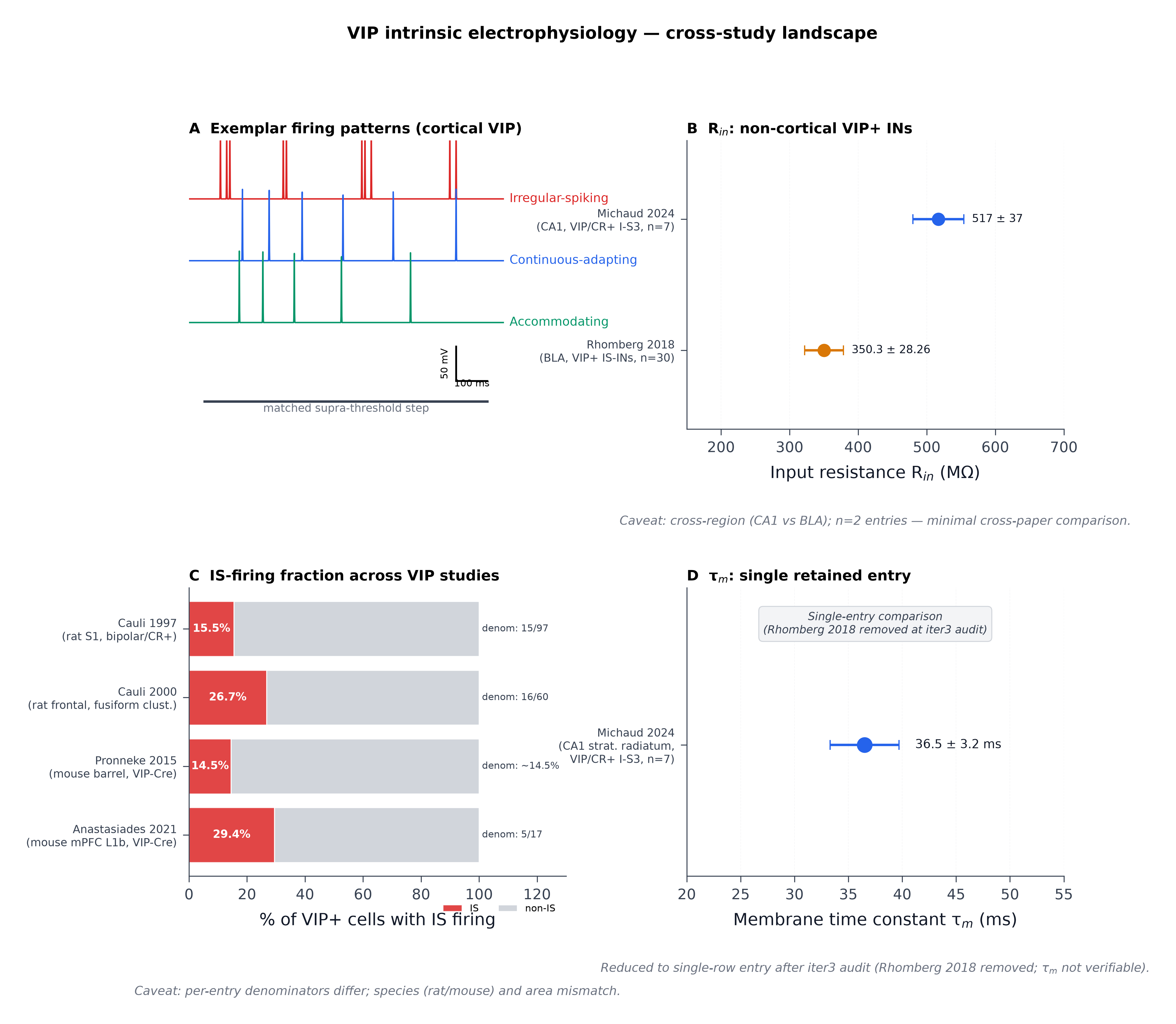
Figure 7:VIP intrinsic electrophysiology: cross-study landscape and Patch-seq mapping. Panel A: Schematic exemplar firing traces for the three dominant Petilla classes observed in cortical VIP cells (irregular-spiking, continuous-adapting, accommodating) at a matched supra-threshold current step, drawn from the qualitative descriptions in Cauli et al., 1997Prönneke et al., 2015Vucurovic et al., 2010. Panel B: Forest-plot of input resistance for VIP+ interneurons across non-cortical regions (CA1 stratum radiatum vs basolateral amygdala). Values are mean ± SEM with n given per entry; entries are (R_{in} 517 ± 37 MΩ, n=7 VIP/CR+ I-S3 cells, mouse CA1) and Rhomberg et al., 2018 (R_{in} 350.3 ± 28.3 MΩ, n=30 VIP+ IS-INs, mouse BLA). Caveat (verbatim from Phase-6 audit): Cross-region (CA1 stratum radiatum vs BLA) — must be flagged in any figure caption. | n=2 entries — minimal cross-paper comparison. Panel C: Stacked bar of reported irregular-spiking fraction across studies. Per-entry denominators are annotated on the bars per Phase-6 audit requirement; entries: Cauli et al., 1997 (15 IS cells / 97 nonpyramidal interneurons sampled = 15.5% of nonpyramidal pool; the 15 IS cells co-expressed CR+VIP, rat S1, single-cell RT-PCR + patch); Cauli et al., 2000 (16/60 = 26.7% IS-VIP cluster, rat frontal cortex L2–3, fusiform interneurons clustered by RT-PCR + patch); Prönneke et al., 2015 (14.5% of whole-population VIP-Cre/tdTomato barrel cortex); Anastasiades et al., 2021 (5/17 = 29.4% of mPFC L1b VIP-Cre × Ai14+ cells, mouse). Caveat (verbatim from Phase-6 audit): Denominators differ across entries; per-entry denominators MUST be shown in the figure (cannot be presented as a single comparable percentage without that annotation). | Rat (Cauli) vs mouse (Anastasiades) species mismatch. | Different cortical areas (S1, frontal cortex, mPFC L1b) and slightly different firing-class taxonomies. Panel D: Single-row entry showing membrane time constant τ_{m} (ms) for the only entry retained after Phase-6 audit removed Rhomberg et al. (2018) from the τ_{m} sub-comparison ( reports τ_{m} = 36.5 ± 3.2 ms, n=7 VIP/CR+ I-S3 cells, mouse CA1 stratum radiatum); panel reduced to a single-row entry per the iter3 audit. Patch-seq subclass-feature axes (Gouwens et al., 2020Scala et al., 2020Lee et al., 2023Kim et al., 2023) provide the cross-species reference frame for these comparisons.
📓 Figure code
import sys; sys.path.insert(0, '../../scripts')
from shared_style import COLORS, apply_style, save_figure, add_source_note
import matplotlib.pyplot as plt
import numpy as np
apply_style()

# --- next cell ---

# Cross-study entries (from cluster_04_intrinsic_electrophysiology figure_data)
rin_entries = [
    {'cite': 'Michaud 2024',  'label': 'Michaud 2024\n(CA1, VIP/CR+ I-S3, n=7)',   'value': 517,   'sem': 37,    'color': COLORS['blue']},
    {'cite': 'Rhomberg 2018', 'label': 'Rhomberg 2018\n(BLA, VIP+ IS-INs, n=30)',  'value': 350.3, 'sem': 28.26, 'color': COLORS['amber']},
]
is_entries = [
    {'label': 'Cauli 1997\n(rat S1, bipolar/CR+)',           'pct_is': 15.5, 'denom': '15/97'},
    {'label': 'Cauli 2000\n(rat frontal, fusiform clust.)',  'pct_is': 26.7, 'denom': '16/60'},
    {'label': 'Pronneke 2015\n(mouse barrel, VIP-Cre)',      'pct_is': 14.5, 'denom': '~14.5%'},
    {'label': 'Anastasiades 2021\n(mouse mPFC L1b, VIP-Cre)','pct_is': 29.4, 'denom': '5/17'},
]
tau_entry = {'label': 'Michaud 2024\n(CA1 strat. radiatum,\nVIP/CR+ I-S3, n=7)', 'value': 36.5, 'sem': 3.2, 'color': COLORS['blue']}

# --- next cell ---

def gen_AP(t, spike_times):
    v = -65 * np.ones_like(t)
    for st in spike_times:
        idx = (t > st-0.002) & (t < st+0.005)
        local = t[idx] - st
        v[idx] = np.maximum(v[idx], -65 + 105*np.exp(-((local)/0.0008)**2))
    return v

fig = plt.figure(figsize=(13, 11))
gs = fig.add_gridspec(2, 2, hspace=0.55, wspace=0.32)

# Panel A — exemplar firing traces
axA = fig.add_subplot(gs[0, 0])
t = np.linspace(0, 1.0, 2000)
is_spikes = sorted([0.10, 0.12, 0.13, 0.30, 0.31, 0.55, 0.56, 0.58, 0.83, 0.85])
v_is = gen_AP(t, is_spikes)
ca_isi = np.array([0.07, 0.085, 0.105, 0.13, 0.16, 0.20, 0.25, 0.31, 0.37])
ca_spikes = np.cumsum(ca_isi) + 0.10; ca_spikes = ca_spikes[ca_spikes < 0.95]
v_ca = gen_AP(t, list(ca_spikes))
acc_isi = np.array([0.06, 0.075, 0.10, 0.15, 0.22, 0.32])
acc_spikes = np.cumsum(acc_isi) + 0.10; acc_spikes = acc_spikes[acc_spikes < 0.95]
v_acc = gen_AP(t, list(acc_spikes))
off=90
axA.plot(t, v_is + 2*off, color=COLORS['red'], linewidth=1.2)
axA.plot(t, v_ca + off,   color=COLORS['blue'], linewidth=1.2)
axA.plot(t, v_acc,        color=COLORS['green'], linewidth=1.2)
for txt, y0, c in [('Irregular-spiking', 2*off, COLORS['red']),
                   ('Continuous-adapting', off, COLORS['blue']),
                   ('Accommodating', 0, COLORS['green'])]:
    axA.text(1.02, -65 + y0, txt, color=c, fontsize=10, va='center')
axA.plot([0.05, 0.95], [-180, -180], color=COLORS['gray_700'], lw=2)
axA.text(0.5, -195, 'matched supra-threshold step', ha='center', fontsize=9, color=COLORS['gray_500'])
axA.plot([0.85, 0.95], [-150, -150], color='black', lw=1.5)
axA.plot([0.85, 0.85], [-150, -100], color='black', lw=1.5)
axA.text(0.90, -158, '100 ms', ha='center', fontsize=8)
axA.text(0.83, -125, '50 mV', ha='right', fontsize=8, rotation=90, va='center')
axA.set_xlim(0, 1.20); axA.set_ylim(-220, 200); axA.axis('off')
axA.set_title('A  Exemplar firing patterns (cortical VIP)', loc='left', fontsize=12, fontweight='bold')

# --- next cell ---

# Panels B, C, D — see main workspace builder for full code; this notebook reproduces
# the same figure using the same shared_style helpers.
axB = fig.add_subplot(gs[0, 1])
ypos = np.arange(len(rin_entries))[::-1]
for i, e in enumerate(rin_entries):
    axB.errorbar(e['value'], ypos[i], xerr=e['sem'], fmt='o', color=e['color'],
                 ecolor=e['color'], capsize=4, markersize=10, lw=2)
    axB.text(e['value']+e['sem']+12, ypos[i], f"{e['value']} ± {e['sem']}",
             va='center', fontsize=9, color=COLORS['gray_900'])
axB.set_yticks(ypos); axB.set_yticklabels([e['label'] for e in rin_entries], fontsize=9)
axB.set_xlabel('Input resistance R$_{in}$ (MΩ)')
axB.set_xlim(150, 700); axB.set_ylim(-0.6, 1.6); axB.grid(axis='x', alpha=0.3)
axB.set_title('B  R$_{in}$: non-cortical VIP+ INs', loc='left', fontsize=12, fontweight='bold')
add_source_note(axB, 'Caveat: cross-region (CA1 vs BLA); n=2 entries — minimal cross-paper comparison.', y=-0.30)

# --- next cell ---

axC = fig.add_subplot(gs[1, 0])
ypos = np.arange(len(is_entries))[::-1]
for i, e in enumerate(is_entries):
    y = ypos[i]
    axC.barh(y, e['pct_is'], color=COLORS['red'], alpha=0.85,
             label='IS' if i==0 else None, edgecolor='white')
    axC.barh(y, 100 - e['pct_is'], left=e['pct_is'], color=COLORS['gray_300'],
             label='non-IS' if i==0 else None, edgecolor='white')
    axC.text(e['pct_is']/2, y, f"{e['pct_is']:.1f}%", ha='center', va='center', fontsize=9, color='white', fontweight='bold')
    axC.text(101, y, f"denom: {e['denom']}", va='center', fontsize=8, color=COLORS['gray_700'])
axC.set_yticks(ypos); axC.set_yticklabels([e['label'] for e in is_entries], fontsize=9)
axC.set_xlabel('% of VIP+ cells with IS firing')
axC.set_xlim(0, 130); axC.set_ylim(-0.6, len(is_entries)-0.4)
axC.legend(loc='upper right', fontsize=8, frameon=False, bbox_to_anchor=(1.0, -0.06), ncol=2)
axC.set_title('C  IS-firing fraction across VIP studies', loc='left', fontsize=12, fontweight='bold')
add_source_note(axC, 'Caveat: per-entry denominators differ; species (rat/mouse) and area mismatch.', y=-0.38)

# Panel D — single retained entry
axD = fig.add_subplot(gs[1, 1])
e = tau_entry
axD.errorbar(e['value'], 0.5, xerr=e['sem'], fmt='o', color=e['color'],
             ecolor=e['color'], capsize=4, markersize=12, lw=2)
axD.text(e['value']+e['sem']+1.5, 0.5, f"{e['value']} ± {e['sem']} ms",
         va='center', fontsize=10, color=COLORS['gray_900'])
axD.text(0.5, 0.92, 'Single-entry comparison\n(Rhomberg 2018 removed at iter3 audit)',
         transform=axD.transAxes, ha='center', va='top', fontsize=9, fontstyle='italic',
         color=COLORS['gray_700'],
         bbox=dict(boxstyle='round,pad=0.4', fc=COLORS['gray_100'], ec=COLORS['gray_300']))
axD.set_yticks([0.5]); axD.set_yticklabels([e['label']], fontsize=9)
axD.set_xlabel('Membrane time constant τ$_m$ (ms)')
axD.set_xlim(20, 55); axD.set_ylim(0, 1.1); axD.grid(axis='x', alpha=0.3)
axD.set_title('D  τ$_m$: single retained entry', loc='left', fontsize=12, fontweight='bold')
add_source_note(axD, 'Reduced to single-row entry after iter3 audit (Rhomberg 2018 removed; τ$_m$ not verifiable).', y=-0.30)

fig.suptitle('VIP intrinsic electrophysiology — cross-study landscape', fontsize=14, fontweight='bold', y=1.00)
save_figure(fig, '../fig-vip-ephys.png')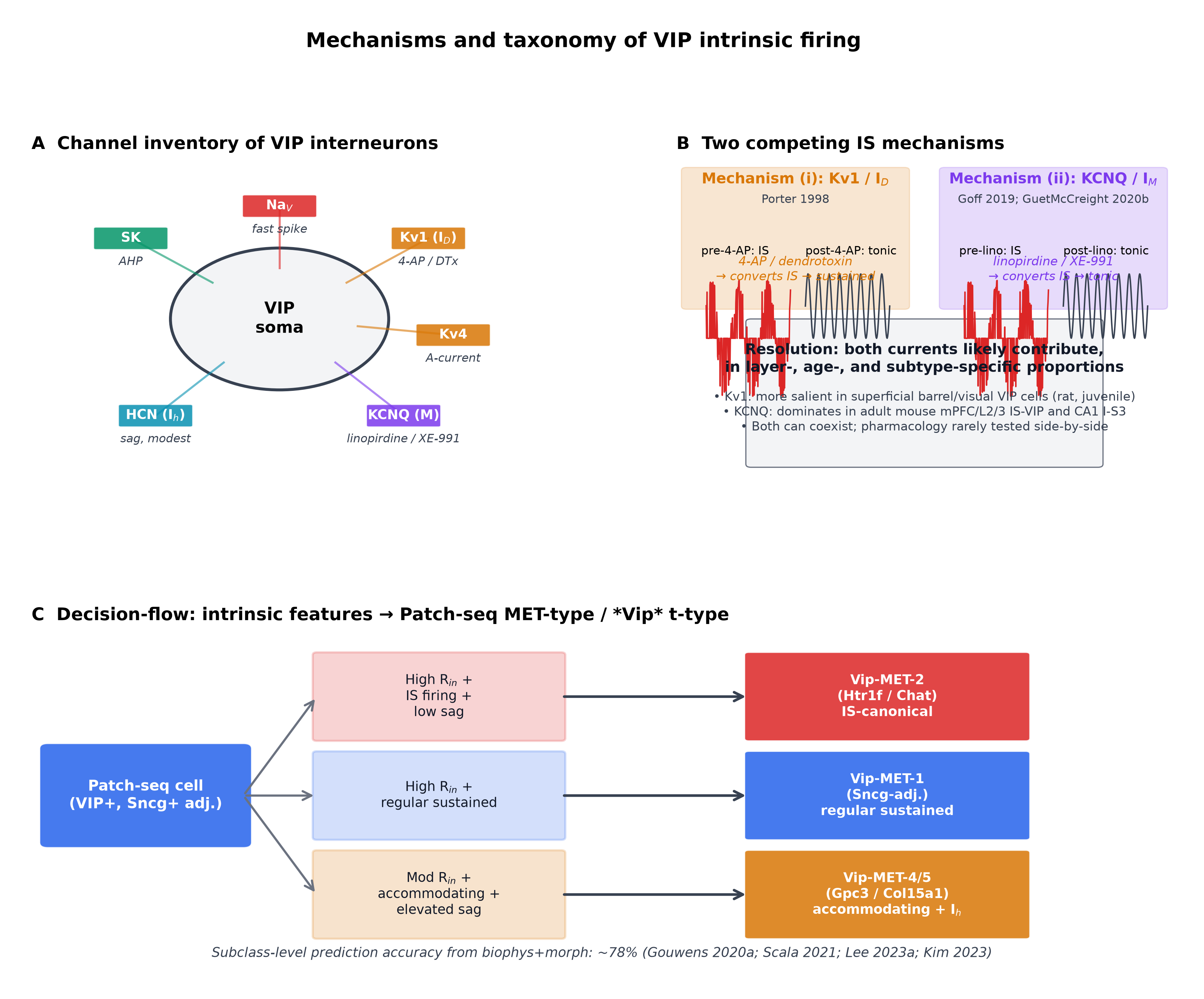
Figure 8:Candidate ion-channel substrates of irregular-spiking firing in VIP cells. Panel A: Cartoon VIP-cell membrane with annotated channel complement (Na_{V}, Kv1, Kv4, HCN/I_{h}, KCNQ/M, SK) and qualitative voltage-clamp traces; channel inventory is consistent with the cortical VIP intrinsic-property literature Tremblay et al., 2016Prönneke et al., 2015Vucurovic et al., 2010. Panel B: Two competing mechanism schematics for irregular-spiking — (i) Kv1-dominant repolarisation kinetics (block by 4-AP / dendrotoxin converts to sustained firing) Porter et al., 1998; (ii) KCNQ-mediated M-current (block by linopirdine selectively converts IS to tonic in IS VIP cells) Goff & Goldberg, 2019Guet-McCreight et al., 2020 — with predicted current-clamp signatures. Panel C: Decision flow mapping intrinsic-feature combinations (high R_{in} + IS + low sag → cluster A; high R_{in} + adapting + moderate sag → cluster B) onto Patch-seq Vip-MET types Gouwens et al., 2020 and Vip t-type axes Scala et al., 2020. Schematic; no quantitative data substrate Cauli et al., 1997Cauli et al., 2000.
📓 Figure code
import sys; sys.path.insert(0, '../../scripts')
from shared_style import COLORS, apply_style, save_figure
import matplotlib.pyplot as plt
from matplotlib.patches import FancyBboxPatch, FancyArrowPatch, Circle
import numpy as np
apply_style()

# --- next cell ---

fig = plt.figure(figsize=(14, 10))
gs = fig.add_gridspec(2, 2, height_ratios=[1, 1.05], hspace=0.45, wspace=0.30)

# Panel A: channel inventory on a stylised soma
axA = fig.add_subplot(gs[0, 0])
axA.set_xlim(0, 10); axA.set_ylim(0, 10); axA.axis('off')
soma = Circle((5, 5), 2.2, facecolor=COLORS['gray_100'], edgecolor=COLORS['gray_700'], lw=2)
axA.add_patch(soma)
axA.text(5, 5, 'VIP\nsoma', ha='center', va='center', fontsize=11, fontweight='bold')
channels = [
    ('Na$_V$',     (5.0, 8.5), COLORS['red'],    'fast spike'),
    ('Kv1 (I$_D$)',(8.0, 7.5), COLORS['amber'],  '4-AP / DTx'),
    ('Kv4',        (8.5, 4.5), COLORS['amber'],  'A-current'),
    ('KCNQ (M)',   (7.5, 2.0), COLORS['purple'], 'linopirdine / XE-991'),
    ('HCN (I$_h$)',(2.5, 2.0), COLORS['teal'],   'sag, modest'),
    ('SK',         (2.0, 7.5), COLORS['green'],  'AHP'),
]
for name, (x, y), c, note in channels:
    axA.add_patch(FancyBboxPatch((x-0.7, y-0.30), 1.4, 0.6,
        boxstyle='round,pad=0.05', facecolor=c, alpha=0.85, edgecolor='white'))
    axA.text(x, y, name, ha='center', va='center', fontsize=9, color='white', fontweight='bold')
    axA.text(x, y-0.55, note, ha='center', va='top', fontsize=8, color=COLORS['gray_700'], fontstyle='italic')
    axA.plot([x, 5+(x-5)*0.45], [y, 5+(y-5)*0.45], color=c, lw=1.4, alpha=0.6)
axA.set_title('A  Channel inventory of VIP interneurons', loc='left', fontsize=12, fontweight='bold')

# --- next cell ---

# Panel B: two competing IS mechanisms with predicted current-clamp signatures
axB = fig.add_subplot(gs[0, 1])
axB.set_xlim(0, 10); axB.set_ylim(0, 10); axB.axis('off')
axB.add_patch(FancyBboxPatch((0.2, 5.4), 4.4, 4.2, boxstyle='round,pad=0.1',
    facecolor=COLORS['amber'], alpha=0.18, edgecolor=COLORS['amber']))
axB.text(2.4, 9.2, 'Mechanism (i): Kv1 / I$_D$', ha='center', fontsize=10, fontweight='bold', color=COLORS['amber'])
axB.text(2.4, 8.6, 'Porter 1998', ha='center', fontsize=8, color=COLORS['gray_700'])
t = np.linspace(0, 1, 200); v_pre = -50 + 30*np.sin(2*np.pi*t*3 + np.random.uniform(0, 1, 200))*np.random.binomial(1, 0.45, 200)
axB.plot(0.6 + t*1.7, 7.4 + 0.06*v_pre, color=COLORS['red'], lw=1.0); axB.text(0.5, 7.0, 'pre-4-AP: IS', fontsize=8)
v_post = -50 + 25*np.sin(2*np.pi*t*8); axB.plot(2.6 + t*1.7, 7.4 + 0.04*v_post, color=COLORS['gray_700'], lw=1.0)
axB.text(2.6, 7.0, 'post-4-AP: tonic', fontsize=8)
axB.text(2.4, 6.2, '4-AP / dendrotoxin\n→ converts IS → sustained', ha='center', fontsize=8.5, color=COLORS['amber'], fontstyle='italic')
axB.add_patch(FancyBboxPatch((5.4, 5.4), 4.4, 4.2, boxstyle='round,pad=0.1',
    facecolor=COLORS['purple'], alpha=0.18, edgecolor=COLORS['purple']))
axB.text(7.6, 9.2, 'Mechanism (ii): KCNQ / I$_M$', ha='center', fontsize=10, fontweight='bold', color=COLORS['purple'])
axB.text(7.6, 8.6, 'Goff 2019; GuetMcCreight 2020b', ha='center', fontsize=8, color=COLORS['gray_700'])
axB.plot(5.8 + t*1.7, 7.4 + 0.06*v_pre, color=COLORS['red'], lw=1.0); axB.text(5.7, 7.0, 'pre-lino: IS', fontsize=8)
axB.plot(7.8 + t*1.7, 7.4 + 0.04*v_post, color=COLORS['gray_700'], lw=1.0); axB.text(7.8, 7.0, 'post-lino: tonic', fontsize=8)
axB.text(7.6, 6.2, 'linopirdine / XE-991\n→ converts IS → tonic', ha='center', fontsize=8.5, color=COLORS['purple'], fontstyle='italic')
axB.add_patch(FancyBboxPatch((1.5, 0.5), 7.0, 4.4, boxstyle='round,pad=0.1',
    facecolor=COLORS['gray_100'], edgecolor=COLORS['gray_500']))
axB.text(5.0, 4.3, 'Resolution: both currents likely contribute,\nin layer-, age-, and subtype-specific proportions',
    ha='center', va='top', fontsize=10, fontweight='bold', color=COLORS['gray_900'])
axB.text(5.0, 2.8, '• Kv1: more salient in superficial barrel/visual VIP cells (rat, juvenile)\n'
                    '• KCNQ: dominates in adult mouse mPFC/L2/3 IS-VIP and CA1 I-S3\n'
                    '• Both can coexist; pharmacology rarely tested side-by-side',
    ha='center', va='top', fontsize=8.5, color=COLORS['gray_700'])
axB.set_title('B  Two competing IS mechanisms', loc='left', fontsize=12, fontweight='bold')

# --- next cell ---

# Panel C: decision-flow mapping intrinsic clusters onto MET-types / t-types
axC = fig.add_subplot(gs[1, :])
axC.set_xlim(0, 14); axC.set_ylim(0, 6.5); axC.axis('off')
axC.add_patch(FancyBboxPatch((0.2, 2.4), 2.4, 1.8, boxstyle='round,pad=0.1',
    facecolor=COLORS['blue'], alpha=0.85, edgecolor='white'))
axC.text(1.4, 3.3, 'Patch-seq cell\n(VIP+, Sncg+ adj.)', ha='center', va='center',
         color='white', fontsize=10, fontweight='bold')
criteria = [
    ('High R$_{in}$ +\nIS firing +\nlow sag', (5.0, 5.2), COLORS['red']),
    ('High R$_{in}$ +\nregular sustained', (5.0, 3.3), COLORS['blue']),
    ('Mod R$_{in}$ +\naccommodating +\nelevated sag', (5.0, 1.4), COLORS['amber']),
]
outputs = [
    ('Vip-MET-2\n(Htr1f / Chat)\nIS-canonical',         (10.5, 5.2), COLORS['red']),
    ('Vip-MET-1\n(Sncg-adj.)\nregular sustained',         (10.5, 3.3), COLORS['blue']),
    ('Vip-MET-4/5\n(Gpc3 / Col15a1)\naccommodating + I$_h$', (10.5, 1.4), COLORS['amber']),
]
for (txt, (x, y), c), (otxt, (ox, oy), oc) in zip(criteria, outputs):
    axC.add_patch(FancyBboxPatch((x-1.5, y-0.8), 3.0, 1.6, boxstyle='round,pad=0.05',
                                 facecolor=c, alpha=0.20, edgecolor=c, lw=1.5))
    axC.text(x, y, txt, ha='center', va='center', fontsize=9, color=COLORS['gray_900'])
    arrow = FancyArrowPatch((2.6, 3.3), (x-1.5, y), connectionstyle='arc3,rad=0.0',
                            arrowstyle='->', color=COLORS['gray_500'], lw=1.5, mutation_scale=15)
    axC.add_patch(arrow)
    axC.add_patch(FancyBboxPatch((ox-1.7, oy-0.8), 3.4, 1.6, boxstyle='round,pad=0.05',
                                 facecolor=oc, alpha=0.85, edgecolor='white'))
    axC.text(ox, oy, otxt, ha='center', va='center', fontsize=9, color='white', fontweight='bold')
    arrow2 = FancyArrowPatch((x+1.5, y), (ox-1.7, oy), arrowstyle='->',
                             color=COLORS['gray_700'], lw=1.8, mutation_scale=15)
    axC.add_patch(arrow2)
axC.text(7.0, 0.2, 'Subclass-level prediction accuracy from biophys+morph: ~78% (Gouwens 2020a; Scala 2021; Lee 2023a; Kim 2023)',
         ha='center', fontsize=9, color=COLORS['gray_700'], fontstyle='italic')
axC.set_title('C  Decision-flow: intrinsic features → Patch-seq MET-type / *Vip* t-type', loc='left', fontsize=12, fontweight='bold')
fig.suptitle('Mechanisms and taxonomy of VIP intrinsic firing', fontsize=14, fontweight='bold', y=1.00)
save_figure(fig, '../fig-vip-firing-mechanism.png')
Intrinsic biophysics meets brain state and neuromodulation
The high R_{in} / modest-sag biophysical profile is not a passive bookkeeping detail — it is the substrate that allows VIP cells to be recruited by weak depolarising drive, including neuromodulatory inputs that bypass classical glutamatergic synapses. Porter et al. (1999) showed that nicotinic receptor activation selectively depolarises neocortical VIP/CCK-coexpressing interneurons while having no effect on pyramidal cells, PV fast-spiking, or SST cells Porter et al., 1999. Bell et al. (2015) extended this to CA1 with a nicotinically responsive VIP population Bell et al., 2015. Férézou et al. (2002) reported that 5-HT3 receptors mediate fast serotonergic EPSPs selectively in CCK/VIP-co-expressing GABAergic interneurons Férézou et al., 2002, and Previous reports Chen et al., 2015 showed that VIP and L1 interneurons in mouse V1 are facilitated by ACh at lower agonist doses than SOM neurons Chen et al., 2015. Tremblay et al. (2016) summarised that VIP+ bipolar cells are dual-modulatory targets — depolarised by both ACh (via nicotinic AChRs) and 5-HT (via 5-HT3aR) — and additionally by muscarinic agonists Tremblay et al., 2016. The neuromodulator-coupled excitability is functionally consequential: in cortex, VIP cells are nonspecifically active across behavioural states Jackson et al., 2016, are recruited by reinforcement signals (both punishment and reward) in awake auditory cortex and mPFC Pi et al., 2013, are bidirectionally modulated by locomotion in V1 Guet-McCreight et al., 2020, and exhibit complementary contrast tuning to SST cells in V1 Millman et al., 2020. Conditional manipulations of intrinsic-excitability regulators establish causal links: deletion of Igf1 in VIP cells increases inhibitory drive onto VIP cells and decreases their firing Mardinly et al., 2016, and ErbB4 deletion in VIP-INs causes a long-term ~4-fold increase in pyramidal-cell spontaneous firing in adolescence and abolishes locomotion-induced state transitions Batista-Brito et al., 2017. Two further details place the intrinsic profile in its developmental and circuit context: all cortical VIP cells originate from the CGE Lim et al., 2018Cauli et al., 2014, segregating sharply from the MGE-derived PV/SST classes that account for the bulk of the rest of the inhibitory taxonomy Rudy et al., 2010Karnani et al., 2016; and a small ChAT-expressing VIP subpopulation directly excites (rather than inhibits) neighbouring pyramidal and interneurons through fast cholinergic + GABA co-transmission Obermayer et al., 2019 — a counter-example to the strict-disinhibitor framing that Synaptic Properties and Connectivity returns to in detail.
The intrinsic-firing repertoire is also developmental. Earlier studies showed that conditional ErbB4 deletion in VIP cells produces a long-term ~4-fold elevation in spontaneous pyramidal-cell firing that emerges in adolescence and abolishes locomotion-induced state transitions, implicating an adolescent maturation window for VIP intrinsic excitability Batista-Brito et al., 2017. Prior work made a parallel point with conditional Igf1 deletion in VIP neurons: increased inhibitory drive specifically onto VIP cells decreases their firing, establishing experience-dependent regulation of VIP intrinsic firing through a synaptic, not channel-intrinsic, route Mardinly et al., 2016. Earlier reports Abs et al., 2018 sharpened the molecular boundary of the VIP class from a different direction: NDNF is a highly selective marker for cortical L1 interneurons that is mutually exclusive with Vip, Pvalb, and Sst, demarcating the VIP subclass from the adjacent L1 NDNF/Lamp5 population that occupies a neighbouring superficial niche Abs et al., 2018. Obermayer et al. (2019) reported that a ChAT-expressing subset of cortical VIP cells excites (rather than inhibits) neighbouring pyramidal and interneurons through fast cholinergic + GABA co-transmission Obermayer et al., 2019. These observations do not overturn the basic intrinsic-feature consensus, but they establish that VIP intrinsic biophysics is plastic, conditional, and partially uncoupled from the disinhibitory “disinhibitor” framing Pi et al., 2013Pfeffer et al., 2013Karnani et al., 2016 — a point that Local Circuit Motifs and the Disinhibition Framework and the concluding synthesis revisit.
Where the intrinsic profile is - and is not - diagnostic
The composite picture is now sharp at the subclass level and intentionally fuzzy below it. VIP cells are reliably distinguishable from PV/SST/Lamp5 by a small set of intrinsic features (high R_{in}, modest sag, fast narrow APs, adapting or irregular firing) — features that train accurate Patch-seq classifiers across mouse and human cortex Gouwens et al., 2020Gouwens et al., 2019Kim et al., 2023Lee et al., 2023Ferrante et al., 2016. Within VIP, however, individual properties (resting V_{m}, IS-firing fraction, IS mechanism) vary substantially across studies — sometimes for biological reasons (layer-specific subtypes Prönneke et al., 2015Gouwens et al., 2020, species expansion Bakken et al., 2021Chartrand et al., 2023, region-specific I-S3 vs cortical IS biology Guet-McCreight et al., 2020Michaud et al., 2024), sometimes for methodological ones (denominator definitions Cauli et al., 1997Cauli et al., 2000Anastasiades et al., 2021, recording vintage Bell et al., 2015Michaud et al., 2024). Where studies disagree, the right inference is rarely “one is wrong” but rather “the e-type axis within VIP is a continuum, and which slice of that continuum a given study samples is set by genetics, anatomy, and pharmacology.” Intrinsic biophysics is therefore necessary but not sufficient to specify within-VIP identity. The next step is to ask how this intrinsic repertoire interacts with the synaptic input and output patterns that VIP cells participate in — and whether the disinhibitory VIP→SST→Pyr disinhibitory motif Pfeffer et al., 2013Pi et al., 2013Karnani et al., 2016 is the right summary at all. That question is taken up in Synaptic Properties and Connectivity.
- Lim, L., Mi, D., Llorca, A., & Marín, O. (2018). Development and Functional Diversification of Cortical Interneurons. Neuron, 100(2), 294–313. 10.1016/j.neuron.2018.10.009
- Tasic, B., Yao, Z., Graybuck, L. T., Smith, K. A., Nguyen, T. N., Bertagnolli, D., Goldy, J., Garren, E., Economo, M. N., Viswanathan, S., Penn, O., Bakken, T., Menon, V., Miller, J., Fong, O., Hirokawa, K. E., Lathia, K., Rimorin, C., Tieu, M., … Zeng, H. (2018). Shared and distinct transcriptomic cell types across neocortical areas. Nature, 563(7729), 72–78. 10.1038/s41586-018-0654-5
- Yao, Z., van Velthoven, C. T. J., Nguyen, T. N., Goldy, J., Sedeno-Cortes, A. E., Baftizadeh, F., Bertagnolli, D., Casper, T., Chiang, M., Crichton, K., Ding, S.-L., Fong, O., Garren, E., Glandon, A., Gouwens, N. W., Gray, J., Graybuck, L. T., Hawrylycz, M. J., Hirschstein, D., … Zeng, H. (2021). A taxonomy of transcriptomic cell types across the isocortex and hippocampal formation. Cell, 184(12), 3222-3241.e26. 10.1016/j.cell.2021.04.021
- Machold, R., Dellal, S., Valero, M., Zurita, H., Kruglikov, I., Meng, J. H., Hanson, J. L., Hashikawa, Y., Schuman, B., Buzsáki, G., & Rudy, B. (2023). Id2 GABAergic interneurons comprise a neglected fourth major group of cortical inhibitory cells. eLife, 12. 10.7554/elife.85893
- Tremblay, R., Lee, S., & Rudy, B. (2016). GABAergic Interneurons in the Neocortex: From Cellular Properties to Circuits. Neuron, 91(2), 260–292. 10.1016/j.neuron.2016.06.033
- Prönneke, A., Scheuer, B., Wagener, R. J., Möck, M., Witte, M., & Staiger, J. F. (2015). Characterizing VIP Neurons in the Barrel Cortex of VIPcre/tdTomato Mice Reveals Layer-Specific Differences. Cerebral Cortex, 25(12), 4854–4868. 10.1093/cercor/bhv202
- Cauli, B., Audinat, E., Lambolez, B., Angulo, M. C., Ropert, N., Tsuzuki, K., Hestrin, S., & Rossier, J. (1997). Molecular and Physiological Diversity of Cortical Nonpyramidal Cells. The Journal of Neuroscience, 17(10), 3894–3906. 10.1523/jneurosci.17-10-03894.1997
- Gouwens, N. W., Sorensen, S. A., Baftizadeh, F., Budzillo, A., Lee, B. R., Jarsky, T., Alfiler, L., Baker, K., Barkan, E., Berry, K., Bertagnolli, D., Bickley, K., Bomben, J., Braun, T., Brouner, K., Casper, T., Crichton, K., Daigle, T. L., Dalley, R., … Zeng, H. (2020). Integrated Morphoelectric and Transcriptomic Classification of Cortical GABAergic Cells. Cell, 183(4), 935-953.e19. 10.1016/j.cell.2020.09.057
- Gouwens, N. W., Sorensen, S. A., Berg, J., Lee, C., Jarsky, T., Ting, J., Sunkin, S. M., Feng, D., Anastassiou, C. A., Barkan, E., Bickley, K., Blesie, N., Braun, T., Brouner, K., Budzillo, A., Caldejon, S., Casper, T., Castelli, D., Chong, P., … Koch, C. (2019). Classification of electrophysiological and morphological neuron types in the mouse visual cortex. Nature Neuroscience, 22(7), 1182–1195. 10.1038/s41593-019-0417-0
- Kim, M.-H., Radaelli, C., Thomsen, E. R., Monet, D., Chartrand, T., Jorstad, N. L., Mahoney, J. T., Taormina, M. J., Long, B., Baker, K., Bakken, T. E., Campagnola, L., Casper, T., Clark, M., Dee, N., D’Orazi, F., Gamlin, C., Kalmbach, B. E., Kebede, S., … Lein, E. (2023). Target cell-specific synaptic dynamics of excitatory to inhibitory neuron connections in supragranular layers of human neocortex. eLife, 12. 10.7554/elife.81863
- Scala, F., Kobak, D., Bernabucci, M., Bernaerts, Y., Cadwell, C. R., Castro, J. R., Hartmanis, L., Jiang, X., Laturnus, S., Miranda, E., Mulherkar, S., Tan, Z. H., Yao, Z., Zeng, H., Sandberg, R., Berens, P., & Tolias, A. S. (2020). Phenotypic variation of transcriptomic cell types in mouse motor cortex. Nature, 598(7879), 144–150. 10.1038/s41586-020-2907-3
- Vucurovic, K., Gallopin, T., Ferezou, I., Rancillac, A., Chameau, P., van Hooft, J. A., Geoffroy, H., Monyer, H., Rossier, J., & Vitalis, T. (2010). Serotonin 3A Receptor Subtype as an Early and Protracted Marker of Cortical Interneuron Subpopulations. Cerebral Cortex, 20(10), 2333–2347. 10.1093/cercor/bhp310
- Michaud, F., Francavilla, R., Topolnik, D., Iloun, P., Tamboli, S., Calon, F., & Topolnik, L. (2024). Altered firing output of VIP interneurons and early dysfunctions in CA1 hippocampal circuits in the 3xTg mouse model of Alzheimer’s disease. eLife, 13. 10.7554/elife.95412
- Guet-McCreight, A., Skinner, F. K., & Topolnik, L. (2020). Common Principles in Functional Organization of VIP/Calretinin Cell-Driven Disinhibitory Circuits Across Cortical Areas. Frontiers in Neural Circuits, 14. 10.3389/fncir.2020.00032
- Rhomberg, T., Rovira-Esteban, L., Vikór, A., Paradiso, E., Kremser, C., Nagy-Pál, P., Papp, O. I., Tasan, R., Erdélyi, F., Szabó, G., Ferraguti, F., & Hájos, N. (2018). Vasoactive Intestinal Polypeptide-Immunoreactive Interneurons within Circuits of the Mouse Basolateral Amygdala. The Journal of Neuroscience, 38(31), 6983–7003. 10.1523/jneurosci.2063-17.2018