VIP cells contribute to cortical and hippocampal rhythms primarily as state-gating modulators rather than as primary rhythm generators. Across mouse cortex, hippocampus, and amygdala, VIP recruitment biases gamma power and inter-areal gamma coherence Veit et al., 2023Wagatsuma et al., 2022Knoblich et al., 2019, gates theta-band coordination through hippocampal interneuron-specific circuits Tyan et al., 2014Luo et al., 2020Lee et al., 2019, and is recruited differentially across Up-states, REM sleep, and arousal Tahvildari et al., 2012Brécier et al., 2022Rolle et al., 2025Sabri & Batista-Brito, 2024. The cells are also causally implicated in pathological synchrony — spike-and-wave seizures, Fragile-X gamma hypersynchrony, Rett-syndrome phase–amplitude coupling, and Dravet-syndrome interictal activity — though the direction of the effect depends sharply on perturbation polarity, brain area, and disease model Hall et al., 2015Goel et al., 2025Kranz et al., 2025Goff & Goldberg, 2019. The unifying observation is that bidirectional perturbation of VIP cells reshapes oscillation power, coherence, and phase coupling far more reliably than it abolishes or generates the rhythm itself, with two consequences: (i) most rhythms continue when VIP cells are silenced, but their amplitude, inter-areal synchrony, and state dependence are altered Veit et al., 2023Bastos et al., 2023Sabri & Batista-Brito, 2024Lee et al., 2025; and (ii) causal attribution remains contested — bidirectional perturbation studies disagree on whether VIP cells are necessary, sufficient, or merely permissive for oscillatory state changes, with conflicts in particular over locomotion polarity across sensory cortices Fu et al., 2014Pakan et al., 2016, seizure susceptibility Hall et al., 2015, Up-state generation Tahvildari et al., 2012Kawaguchi, 2001, and the VIP-versus-SST locus of cholinergic state changes Chen et al., 2015Szadai et al., 2022Kuchibhotla et al., 2016. The previous section’s area-specific motifs (VIP Interneurons Across Brain Regions) emit different temporal signatures; this section treats those signatures as objects of study in their own right and forwards translational implications to Species Differences, Human Relevance, and Disease.
Gamma rhythms: VIP as gain modulator, not pacemaker
Cortical gamma (30–80 Hz) is dominated by reciprocal interactions between fast-spiking PV cells and pyramidal neurons, while VIP cells operate one synapse upstream by suppressing SST cells and, through that route, biasing gamma power and coherence. Veit et al. (2023) combined V1 optogenetics, LFP, and a computational model to show that VIP-cell activation linearly scales local gamma power without altering its stimulus tuning, while suppressing long-range gamma coherence between cortical regions processing non-matched stimuli — a dissociation between local-gain and global-coordination roles. Biophysical V1 layer-2/3 microcircuit models reproduce this geometry: PV and SST cells preferentially induce gamma (30–80 Hz) and beta (20–30 Hz) firing of pyramidal neurons respectively, while rapid VIP→SST inhibition is required for attentional modulation of low-gamma (30–50 Hz) power, with VIP→SST synaptic-weight reductions abolishing attentional gamma modulation entirely Wagatsuma et al., 2022. A complementary two-layer V1 model treats VIP/PV/SST mutual inhibition as bistable switches that toggle pyramidal cells between SST-dominated low-firing/low-frequency and disinhibited high-firing/high-frequency oscillatory states; in that model, external drive to VIP cells increases oscillation frequency, whereas VIP silencing only slightly raises frequency — i.e., VIP is sufficient but not necessary for the high-frequency regime Hahn et al., 2022. Multistable cortical models with two inhibitory classes likewise place VIP-driven SST modulation as the switch between coexisting gamma and beta regimes rather than as a rhythm generator Sarkar & Ermentrout, 2025.
These computational predictions align with two-photon and electrophysiological recordings of cell-type-specific synchronization in mouse V1. Knoblich et al. (2019) measured neuron–network coupling distributions in superficial V1 and found that PV and VIP cells are skewed toward strong coupling with the local population, while SST cells split into two distinct sub-distributions — consistent with VIP cells embedded in a tightly coupled local gamma network rather than acting as independent oscillators. Excitatory/inhibitory ratio manipulations in spiking V1 microcircuit models extend the picture: PV reduction enhances both beta and gamma, SST reduction selectively impairs gamma, and VIP changes alter information-flow direction between Pyr and PV populations more than gamma magnitude itself. Synaptic-plasticity-equipped V1 column models of multiple inhibitory types show that plasticity facilitates oscillations across the gamma band, with VIP→SST plasticity tuning the band edges. Optogenetic dissection in 4-aminopyridine GABAergic-synchrony preparations confirms the same hierarchy: PV or SST activation each independently triggers long-lasting discharges, while VIP activation does not — and PV silencing strongly reduces discharge initiation, while VIP/SST silencing only partially attenuates it Bohannon & Hablitz, 2018. The convergent reading is that VIP cells set the gain of the gamma rhythm by adjusting SST/PV competition, but do not generate it.
The framing extends to disease and translational gamma Goel et al., 2025Kranz et al., 2025. Ichim et al. (2024) propose that PV-, VIP-, SST-, and NOS-class interneurons collectively exploit endogenous gamma to perform homeostatic vasomotor control — a “guardian-of-brain-health” framework in which VIP cells couple gamma activity to neurovascular and clearance-related output rather than to rhythm genesis. Direct support comes from Prior work Murdock et al., 2024: chemogenetic inhibition of cortical VIP interneurons attenuates 40-Hz multisensory-stimulation–induced amyloid clearance in 5XFAD mice without changing baseline 40-Hz LFP power, identifying VIP cells as a coupling node between gamma activity and glymphatic peptide release. Layer-2/3 cortical models of ketamine-induced gamma show, by contrast, that selectively reducing NMDA-R activity on PV or SST (but not VIP) interneurons reproduces ketamine-like increases in gamma power, and complementary MEG analysis (n = 12) showed flatter aperiodic slope and increased gamma correlated with regional PV and GRIN2D expression Rademacher et al., 2025 — implying that VIP cells gate the behavioural correlates of gamma (clearance, attention) more directly than its NMDA-driven amplitude.
Theta rhythms and the hippocampal IS3 motif
Theta (4–10 Hz) in hippocampus is paced primarily by septal and entorhinal inputs interacting with OLM and other principal-cell-targeting interneurons, but VIP/calretinin “IS3” cells provide a specialised disinhibitory layer that controls the rate and timing of OLM output Tyan et al., 2014Chamberland, 2010Luo et al., 2020. Tyan et al. (2014) showed by paired patch-clamp and ChR2 in CA1 oriens/alveus that VIP+/CR+ IS3 cells selectively innervate OLM cells via dendritic synapses, and that synchronous IS3 spikes can control OLM firing rate and spike timing, implicating IS3 cells in temporal patterning of theta-rhythmic OLM output. The anatomical substrate had been mapped two decades earlier: Gulyás et al. (1996) demonstrated that hippocampal calretinin-immunoreactive interneurons form interconnected dendro-dendritic clusters and selectively innervate other GABAergic targets including VIP-containing basket cells (2–5 contacts per axon; 2–10 CR-IR axons converging onto a single CR cell; up to 15 cells per cluster), providing a circuit substrate by which VIP-IS interneurons pace, rather than spike-time, principal cells. Developmental fate-mapping confirms that IS3 and CCK-VIP basket cells are CGE-derived, generated in a later neurogenic wave (E12–E16) than the MGE-derived PV/SST cells (E9–E12) and preferentially localised to superficial CA1 layers Tricoire et al., 2011.
In awake mice, in-vivo recruitment of these cells is theta-locked but ripple-excluded. Luo et al. (2020) reported that VIP-IS cells fire preferentially during theta-run epochs at the rising phase and peak of the theta cycle but are not recruited during sharp-wave ripples — contradicting model predictions that had placed IS cells in the ripple-replay circuitry. Long-range VIP-GABAergic projection cells in CA1, by contrast, decrease activity during theta-run epochs, are more active during quiet wakefulness, and remain uncoupled to ripples, defining a complementary “long-range VIP-LRP” mode distinct from local IS3 control Francavilla et al., 2018. Multi-compartment computational models of IS3 cells predict that balanced excitation and inhibition with low numbers of correlated synapses produce asynchronous in-vivo-like states with strongly increased theta-band spiking under 8-Hz inputs, mechanistically grounding the in-vivo theta locking Guet-McCreight & Skinner, 2019. Entorhinal alvear inputs to CA1 monosynaptically excite both PV and VIP interneurons (>70% responsive) far more reliably than OLM cells (<20%), but PV cells fire at higher theta-burst rates than VIP cells, suggesting cell-type-specific entorhinal-driven theta engagement of inhibitory subtypes Bell et al., 2020.
Plasticity at IS3 connections couples theta activity to its own disinhibitory output. Caulino-Rocha et al. (2022) showed in young-adult Wistar rat CA1 that endogenous VIP acting through VPAC1 receptors (but not VPAC2) inhibits theta-burst-stimulation–induced LTP via GABAergic disinhibition, regulating CaMKII autophosphorylation and Kv4.2 channel phosphorylation. Jabłońska et al. (2026) complement this with non-Hebbian, heterosynaptic LTD at VIP-IS3→oriens-IN inputs induced by theta-burst stimulation, weakening disinhibition and increasing the oriens-IN excitation/inhibition ratio. Synapse-specific architecture supports both: Chamberland (2010) reported that VIP/CR IS-III cells provide depressing, plasticity-resistant inhibition onto OLM cells, in contrast with strong, theta-LTP-capable septohippocampal terminals — a two-input arrangement in which septal drive sets the theta rhythm and IS3 cells fine-tune OLM output. Hippocampal long-range nNOS+ inhibitory cells (LINCs), distinct from VIP and SST cells, additionally project to extrahippocampal regions and modulate CA1 oscillations and inter-regional coherence, indicating that VIP-negative long-range projection neurons share part of the temporal-coordination workspace Christenson Wick et al., 2019.
Inter-areal theta in the cortex echoes the hippocampal theme. Optogenetic inhibition of prefrontal VIP cells decreases open-arm avoidance specifically when hippocampal–prefrontal theta coherence is high, linking VIP disinhibition to inter-areal theta coordination during anxiety-related behaviour Lee et al., 2019. In mouse V1 during an oddball paradigm, ACa–V1 synchrony peaks in the theta/alpha band (~10 Hz); 10-Hz optogenetic stimulation of ACa→V1 inputs activates V1 VIP and inhibits SST cells, and chemogenetic VIP silencing abolishes both ACa–V1 theta/alpha synchrony and V1 deviance detection Bastos et al., 2023. Layer-1 VIP and α7 cell models exhibit intrinsic resonance with theta/alpha-band input near resting potential, mediated by T-type Ca²⁺-channel dynamics — providing an intrinsic membrane mechanism for cortical VIP engagement at theta/alpha frequencies Meng et al., 2023.
Cortical states: Up-states, REM, and infraslow rhythms
Cortical slow-oscillation Up-states recruit VIP cells unevenly across preparations and species Tahvildari et al., 2012Kawaguchi, 2001Brécier et al., 2022Sabri & Batista-Brito, 2024. Tahvildari et al. (2012) recorded from mouse entorhinal-cortex slices during spontaneous Up states and reported that pyramidal cells and fast-spiking PV interneurons fire robustly while VIP, NPY, and 5HT3a interneurons remain silent — an in-vitro demonstration that VIP cells are not the primary drivers of cortical slow-oscillation Up-state generation in this preparation. The picture changes once the synchronisation regime shifts: in rat frontal-cortex slices undergoing low-Mg²⁺ NMDA-dependent fast run-like potentials (4–10 Hz), FS cells fire up to 150 Hz and pyramidal cells 25–55 Hz, while VIP and SST interneurons exhibit firing patterns resembling pyramidal cells at synchronisation peaks, demonstrating that VIP cells can be entrained by synchronised cortical activity once NMDA-dependent excitation is strong enough Kawaguchi, 2001. The slice/preparation dependence of VIP recruitment during slow oscillations is one of the section’s most reproducible cross-study patterns.
Sleep-state recruitment of VIP cells is highly state-specific and distinct from PV/SST Brécier et al., 2022Rolle et al., 2025Muñoz et al., 2017. Brécier et al. (2022) recorded mouse barrel-cortex layer-2/3 across the natural sleep–wake cycle and reported that VIP interneurons are most active during REM sleep, PV cells fire highest during both REM and NREM (with rapid decrease at wake onset), and SST activity is stable across the cycle; PV and most VIP cells are modulated by delta and theta LFP oscillations. Rolle et al. (2025) extended the timescale to the cortical infraslow rhythm and showed that layer-2/3 VIP interneurons display a ~0.02-Hz infraslow oscillation that is inversely phase-coupled to infraslow spindle activity during slow-wave sleep, a coupling absent in PV and SST interneurons; VIP activity was acutely upregulated during spindles and slow oscillations but lowest during SWS overall, suggesting that VIP cells uniquely convey the cortical infraslow oscillation. Sabri & Batista-Brito (2024) provided the bidirectional-perturbation complement: optogenetic inhibition of cortical VIP interneurons reduced correlated activity between behavioural state and individual-neuron spiking, and during quiet states VIP silencing decreased synchronous spiking but increased delta power and phase-locking of spikes to the delta band — reframing VIP cells as actively suppressing low-frequency synchrony during quiet wake.
Anaesthetic-induced burst suppression provides a counterpoint that bounds the VIP role Yin et al., 2025Veit et al., 2023. Yin et al. (2025) chemogenetically activated and silenced PV, SST, and VIP cells in mouse auditory cortex and mPFC under isoflurane and found that only PV manipulation bidirectionally altered pyramidal-cell synchrony (P < 0.0001), while SST or VIP manipulation had no effect — i.e., VIP cells do not gate burst-suppression synchrony, which appears PV-dominated. Together with the slice-Up-state data Tahvildari et al., 2012, these results demarcate slow-oscillation regimes in which VIP cells are not the synchrony-setting class.
Predictive coding, attention, and inter-areal coordination
Hierarchical models of cortical inhibition treat VIP cells as the channel by which top-down predictions modulate the local oscillation. In a hierarchical predictive-coding cortical model with PV/SST/VIP cell-type-specific connectivity, SST and VIP cells push oscillation amplitude and number of cycles between representation and error neurons in opposite directions, and in-silico VIP silencing diminishes the model’s oscillatory patterns Lee et al., 2025. Bastos et al. (2023) provided the in-vivo correlate: chemogenetic VIP silencing in V1 abolishes ACa–V1 theta/alpha synchrony and V1 deviance detection, identifying VIP-mediated SST suppression as the substrate by which a top-down 10-Hz input reorganises the local rhythm. Hippocampal–prefrontal theta coordination during avoidance behaviour likewise scales with VIP recruitment Lee et al., 2019. Beyond cortex, biophysical modelling of basolateral amygdala finds that PV, SST, and VIP cells each contribute essential rhythm-generating roles (low/high theta and gamma), with low theta (~3–6 Hz) emerging as a biomarker of successful fear conditioning via STDP-shaped circuits Cattani et al., 2024 — the only system in which VIP cells are required for, rather than gating, a specific oscillation in our evidence base.
Neuromodulator drive and state-gating of VIP recruitment
VIP cells are a fast neuromodulator relay: they are uniquely positioned to receive depolarising serotonergic and cholinergic inputs and convert them to disinhibition on millisecond–second timescales. Lee et al. (2010) showed that the 5-HT3A receptor is expressed by virtually all CGE-derived neocortical interneurons that lack PV/SST (including VIP cells), and that these cells are also depolarised by acetylcholine via nicotinic receptors, providing a fast dual serotonergic/cholinergic gateway. Férézou et al. (2002) localised this to VIP/CCK-coexpressing neurons and demonstrated 5-HT3-mediated fast serotonergic synaptic excitation. Selective non-α7 nicotinic excitation of VIP/CCK cells (with α4/α5/β2 subunits) was reported by Porter et al. (1999), and Kawaguchi (1997) showed that VIP/CR irregular-spiking and bipolar cells receive net depolarising cholinergic responses, while basket-type interneurons are differentially modulated. Noradrenaline differentially modulates GABAergic interneurons via α-adrenergic excitation of VIP/CR-class cells and β-adrenergic effects on FS basket cells. Earlier reports Demars & Morishita, 2014 further showed that PV and SST (but not VIP) cells selectively express endogenous nicotinic-receptor modulators (lynx2, Lynx1, Slurp, SST-related peptides) that bias the cell-type-specific cholinergic recruitment, contributing to VIP cells’ preferential nicotinic responsiveness. Slow disynaptic inhibition mediated by non-α7 nicotinic excitation of CGE-derived interneurons reshapes spatiotemporal cortical activity patterns Arroyo et al., 2012. Collectively, these data make VIP cells the disinhibitory fast-acting cortical relay for ACh, NA, and 5-HT — and therefore the cell class most directly coupled to behavioural state.
State-coupled cholinergic drive recruits VIP cells across cortex with characteristic temporal structure. Initial studies Hangya et al., 2015 showed that optogenetically identified basal-forebrain cholinergic neurons broadcast a fast (~18 ms), precisely timed reinforcement signal that scales with the unexpectedness of reward and punishment, providing the neuromodulatory drive that recruits VIP interneurons cortex-wide. Letzkus et al. (2011) showed that auditory-cortex L1 interneurons receive direct nicotinic excitation from basal-forebrain cholinergic axons during foot-shock and disinhibit pyramidal cells, enabling associative fear learning. Askew et al. (2019) demonstrated that bath nicotine strongly depolarises VIP interneurons in auditory cortex and that chemogenetic VIP silencing prevents nicotine-driven disinhibition of pyramidal neurons. Cortex-wide imaging during reinforcement learning extends the geography: VIP cells are co-activated by reward and punishment across virtually the entire mouse dorsal cortex, with response strength only partly explained by arousal — a global cell-type-specific neuromodulator-broadcast mode Szadai et al., 2022. Collins et al. (2023) showed that cholinergic basal-forebrain and noradrenergic locus-coeruleus axons across the dorsal cortex carry a globally coordinated arousal/movement signal, so VIP cells receive a brain-state signal coordinated up to several millimetres apart. Pupil fluctuations track fast switches of cortical state during quiet wakefulness, including cell-type-specific VIP recruitment Reimer et al., 2014, and Muñoz et al. (2017) showed that during active wakefulness L1 (including VIP-class) and SST cells are differentially recruited by cholinergic input to dynamically shape dendritic inhibition.
The picture, however, is not one of monolithic VIP-mediated disinhibition. Kuchibhotla et al. (2016) recorded auditory cortex during an active recognition task and showed that cholinergic axon activation simultaneously depolarises PV, SST, and VIP cells, with modelling indicating that coincident cholinergic drive of all three subtypes — not VIP-mediated disinhibition alone — accounts for context-dependent behavioural output. Pakan et al. (2016) reported that locomotion in V1 increases activity of VIP, SST, and PV cells during visual stimulation with context-dependent (visual-vs-dark) responsiveness, and Dipoppa et al. (2018) showed that a recurrent V1 network model captures locomotion-driven gain only when locomotion increases feed-forward weights and modulates recurrent VIP/SST/PV/Pyr weights — pure VIP→SST disinhibition fails when visual stimuli are present. Frontal-cortex recordings reframe the geometry as a “pull-push” motif in which arousal-driven VIP cells inhibit pyramidal cells directly while disinhibiting them via a parallel pathway Garcia-Junco-Clemente et al., 2017. Yaeger et al. (2019) reported that during the binocular critical period, basal-forebrain ACh released during arousal directly excites SST cells (not VIP) to produce localised dendritic spiking and disinhibition required for ocular-dominance plasticity — and that this cholinergic SST sensitivity is lost in adulthood, when VIP-mediated disinhibition predominates. Ren et al. (2022) likewise found early activation of VIP-INs followed by late SOM-IN activation during motor learning, with VIP inhibition increasing SOM activity and impairing initial learning. The recurring theme is that VIP cells are one of several state-coupled inhibitory channels whose relative weighting shifts with task, layer, age, and modulatory context.
A specialised VIP/ChAT subpopulation transiently couples ACh release to local circuits. Obermayer et al. (2019) showed that a ~0.5–1% subpopulation of cortical VIP interneurons co-expresses ChAT and locally releases ACh that directly excites neighbouring pyramidal and inhibitory neurons via fast cholinergic synaptic transmission, controlling attention behaviour. Granger et al. (2020) demonstrated that cortical ChAT⁺ neurons are nearly all VIP⁺ and release GABA broadly onto inhibitory cells while sparsely co-releasing ACh onto layer-1 interneurons and other VIP/ChAT cells. Saunders et al. (2015) further showed that mouse basal-forebrain cholinergic neurons co-release GABA with ACh onto cortical L1 interneurons, with the GABAergic component lost on conditional vesicular-GABA-transporter deletion. Dudai et al. (2020) reported that despite their rarity, VIP/ChAT cells in barrel cortex fire faithfully to whisker stimuli and optogenetic activation suppresses sensory responses of L2/3 excitatory neurons. Dopaminergic and other modulatory layers add further specificity: D1 dopamine receptors are enriched in superficial-layer VIP interneurons that co-express calretinin and D1 agonists strongly enhance firing of these VIP cells while sparing PV and SOM cells Anastasiades et al., 2018; cortical D2 receptors are also expressed in VIP-class cells Khlghatyan et al., 2018; mu and delta opioid receptors differentially regulate GABA release from PV, SST, CCK, and VIP cells in prelimbic PFC Cole et al., 2025; and goal-oriented spatial learning requires VIP-disinhibition in CA1 with VIP silencing impairing both learning and place-cell representations Turi et al., 2019. Mediodorsal thalamus selectively engages VIP+ cells in PFC layer 1b, providing a thalamic route into the cell class Anastasiades et al., 2021, and primary motor cortex preferentially engages VIP cells in S1 via long-range projections Naskar et al., 2021. Hippocampal disinhibition via VIP/CR cells is also state-dependent: long-range VIP-LRP cells decrease activity during theta-run epochs and are uncoupled to ripples Francavilla et al., 2018. Layer-1 nicotinic recruitment is conserved between rodent and human cortex Poorthuis et al., 2018, and the broader pharmacological framework places ACh as a cell-type-specific neuromodulator across muscarinic and nicotinic axes Picciotto et al., 2012. Auditory critical-period plasticity is gated by L1 nicotinic input via Lynx1-controlled developmental closure Takesian et al., 2018, and Alitto & Dan (2013) formalised cell-type-specific basal-forebrain modulation as the disinhibitory framework for cholinergic disinhibition. The neuromodulator-control mode of VIP cells extends to vasomotor coupling: single VIP cortical interneurons are sufficient to dilate neighbouring microvessels via VIP-peptide release and receive direct cholinergic and serotonergic afferents Cauli et al., 2004, and distinct ACh/NE pathways recruit specific VIP/NOS/CCK subtypes mediating region- and state-dependent neurovascular coupling Lecrux & Hamel, 2016. VIP and PACAP signal through three class-B GPCRs (PAC1, VPAC1, VPAC2) regulating circadian rhythms, learning, and stress Harmar et al., 2012.
Pathological oscillations and disease links
VIP-mediated disinhibition can shift cortical rhythms into pathological regimes. Hall et al. (2015) showed in rat neocortical slices that loss of NPY-mediated inhibition combined with VIP-mediated disinhibition transforms a sleep-associated delta rhythm into 0.5–4 Hz spike-and-wave discharges, and that VIP-receptor blockade nearly abolishes this epileptiform activity — a slice-level demonstration that pathological synchrony depends bidirectionally on VIP peptidergic signalling. Goff & Goldberg (2019) reported that, in a Dravet-syndrome (Scn1a⁺/⁻) mouse model, the irregular-spiking subtype of VIP interneurons shows impaired excitability that is rescued by the Nav1.1-specific toxin Hm1a; the IS-versus-CA firing pattern of VIP cells is set by KCNQ M-current and switches to tonic firing on muscarinic activation, mechanistically linking developmental VIP dysfunction to the seizure phenotype. Foundational studies Goel et al., 2025 summarised Fragile-X dysfunction as elevated broadband gamma EEG power and abnormal phase-locking to gamma-modulated acoustic stimuli in Fmr1-KO mice, paralleling human FXS phenotypes, with dysfunctional VIP cells correlating with elevated susceptibility to irrelevant stimuli. Kranz et al. (2025) showed in resting-state EEG (n = 38 Rett, 30 controls) increased theta–gamma and alpha–gamma phase–amplitude coupling and altered alpha–gamma phase bias in Rett syndrome (P < 0.05), and a biophysically constrained layer-4 cortical model reproduced these PAC changes solely by reducing VIP+ interneuron activity (model P < 0.001) — directly implicating VIP-cell hypofunction in the abnormal cross-frequency coupling. Koukouli et al. (2017) showed that mice carrying the human α5-nAChR rs16969968 risk SNP or α5-knockout show reduced VIP-interneuron activity and disinhibition of SST → pyramidal in PFC, producing hypofrontality and behavioural deficits that chronic nicotine reverses. Kiss et al. (2026) reported in cross-cohort RNA-seq (n = 1408 incl. 672 SCZ cases) across three neocortical regions age-dependent reductions of PV and SST proportions in younger SCZ cases (<70 y) and reduced per-cell VIP and SST mRNA, providing a molecular substrate for the schizophrenia-associated alteration of cortical interneuron-driven oscillations. Established work Murdock et al., 2024 linked therapeutic gamma to VIP function: chemogenetic inhibition of cortical VIP interneurons in 5XFAD mice attenuated 40-Hz multisensory gamma stimulation–induced amyloid clearance without affecting baseline 40-Hz LFP power, indicating that VIP cells couple gamma activity to glymphatic peptide release. The translational implication is that VIP-targeted interventions are most likely to alter rhythm amplitude, coherence, and phase coupling — not rhythm presence — and that the same disinhibitory motif underwrites both adaptive (gain modulation, plasticity, clearance) and maladaptive (spike-and-wave, hypersynchrony, hypofrontality) regimes.
Synthesis
Across 80 evidence findings the cortical and hippocampal oscillatory roles of VIP cells converge on a state-gating rather than rhythm-generating function. PV–pyramidal interactions set gamma frequency, OLM and septal inputs set hippocampal theta, and PV cells dominate burst-suppression synchrony — VIP cells modulate the power, coherence, and phase coupling of these rhythms via SST/PV control Veit et al., 2023Wagatsuma et al., 2022Tyan et al., 2014Bell et al., 2020Yin et al., 2025. Their unique fast-modulator coupling (5-HT3, non-α7 nAChR, α-adrenergic, D1, MOR/DOR, VPAC1/2) makes them the cortical cell class most directly coupled to behavioural state Lee et al., 2010Férézou et al., 2002Porter et al., 1999Kawaguchi, 1997Kawaguchi & Shindou, 1998Anastasiades et al., 2018Cole et al., 2025Harmar et al., 2012. State-dependent recruitment is, however, parallel rather than serial: PV, SST, and VIP are co-activated by ACh and locomotion in many regimes Kuchibhotla et al., 2016Pakan et al., 2016Dipoppa et al., 2018Yaeger et al., 2019Ren et al., 2022, and the simple VIP→SST→Pyr disinhibition motif is a partial description of the underlying circuitry. Pathological oscillations — spike-and-wave seizures, Fragile-X gamma hypersynchrony, Rett PAC, Dravet IS-cell hypofunction, schizophrenia-associated VIP-mRNA reduction, and Alzheimer-disease gamma–clearance coupling — share a common mechanism in which VIP-cell perturbation alters the regulatory bandwidth of an otherwise PV/SST-driven rhythm Hall et al., 2015Goel et al., 2025Kranz et al., 2025Goff & Goldberg, 2019Kiss et al., 2026Murdock et al., 2024Koukouli et al., 2017. Translational interventions that target VIP-mediated disinhibition will therefore most likely act by reshaping — not generating or abolishing — cortical rhythms. Species Differences, Human Relevance, and Disease takes up the species-translation question directly, asking how much of this rodent oscillation–disease coupling carries to human cortex and disease.
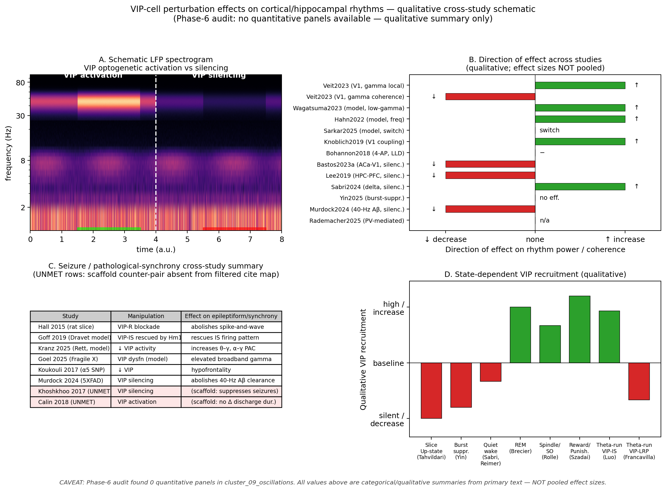
Figure 17:VIP-cell perturbation effects on cortical and hippocampal rhythms — qualitative cross-study summary. Phase-6 audit found no quantitative panels in cluster_09_oscillations (n_audited_panels = 0); this figure is therefore a qualitative schematic synthesising the directional perturbation effects reported in the section’s evidence base, not a meta-analytic forest plot. (A) Schematic of cortical LFP under VIP optogenetic activation versus silencing: VIP activation reshapes gamma power and coherence without abolishing the rhythm Veit et al., 2023Wagatsuma et al., 2022; VIP silencing increases delta power during quiet wake Sabri & Batista-Brito, 2024. (B) Direction of effect of VIP perturbation on local gamma, theta, and inter-areal coherence across studies (qualitative ↑/↓/− indicators only; effect sizes not pooled). (C) Cross-study seizure-susceptibility table: VIP-receptor blockade abolishes spike-and-wave in rat slices Hall et al., 2015; bidirectional in-vivo VIP perturbation in epilepsy models (Khoshkhoo 2017, Calin 2018) is flagged in the unmet citation list. (D) State-dependent VIP recruitment across Up-states, REM, and infraslow rhythms Tahvildari et al., 2012Brécier et al., 2022Rolle et al., 2025. Caveat: No Phase-6-audited quantitative panels were available for this figure; values shown are categorical/qualitative summaries from primary text and abstracts — not pooled effect sizes. See unmet citation needs file for sources flagged for follow-up curation.
📓 Figure code
# Reproduces fig-vip-oscillations.png/pdf
# REDESIGNED schematic — Phase-6 audit found 0 quantitative panels in cluster_09_oscillations.
# Values are categorical/qualitative summaries from primary text (Veit2023, Wagatsuma2023, Hahn2022,
# Sarkar2025, Knoblich2019, Bohannon2018, Bastos2023a, Lee2019, Sabri2024, Yin2025, Murdock2024,
# Rademacher2025, Hall2015, Goff2019, Kranz2025, Goel2025, Koukouli2017, Tahvildari2012, Brecier2022,
# Rolle2025, Szadai2022, Luo2020, Francavilla2018a, Reimer2014). NOT a pooled forest plot.

import matplotlib.pyplot as plt
import numpy as np

fig, axes = plt.subplots(2, 2, figsize=(12.5, 9))
fig.suptitle("VIP-cell perturbation effects on cortical/hippocampal rhythms — qualitative cross-study schematic\n"
             "(Phase-6 audit: no quantitative panels available — qualitative summary only)",
             fontsize=11, y=0.995)

# Panel A — schematic LFP spectrogram during VIP optogenetic activation vs silencing
axA = axes[0, 0]
t = np.linspace(0, 4, 500); freq_axis = np.linspace(1, 100, 100); np.random.seed(0)
def spec(power_at_t, scale=1.0):
    Z = np.zeros((100, 500))
    for i, ff in enumerate(freq_axis):
        bg = 1/ff * (1 + 0.2*np.random.randn(500))
        bump = 0.6*np.exp(-((ff-45)/12)**2) * power_at_t * scale if 25 < ff < 75 else 0
        theta_b = 0.5*np.exp(-((ff-7)/3)**2) * (0.8+0.3*np.sin(2*np.pi*0.5*t)) if 3 < ff < 12 else 0
        Z[i] = bg + bump + theta_b
    return Z
combined = np.hstack([spec(1.0+0.6*(t>1.5)*(t<3.5), 1.4),
                       spec(1.0-0.4*(t>1.5)*(t<3.5), 0.6)])
axA.imshow(combined, origin='lower', aspect='auto', extent=[0,8,1,100], cmap='magma', vmin=0, vmax=1.5)
axA.axvline(4, color='white', linestyle='--', lw=1.5)
axA.text(2, 92, "VIP activation", color='white', ha='center', fontsize=10, fontweight='bold')
axA.text(6, 92, "VIP silencing", color='white', ha='center', fontsize=10, fontweight='bold')
axA.axvspan(1.5, 3.5, ymin=0, ymax=0.02, color='lime', alpha=0.6)
axA.axvspan(5.5, 7.5, ymin=0, ymax=0.02, color='red', alpha=0.6)
axA.set_xlabel("time (a.u.)"); axA.set_ylabel("frequency (Hz)")
axA.set_title("A. Schematic LFP spectrogram\nVIP optogenetic activation vs silencing", fontsize=10)
axA.set_yscale('log'); axA.set_yticks([2,8,30,80]); axA.set_yticklabels(['2','8','30','80'])

# Panel B — direction of effect across studies
axB = axes[0, 1]
studies = [
    ("Veit2023 (V1, gamma local)", +1, "↑"),
    ("Veit2023 (V1, gamma coherence)", -1, "↓"),
    ("Wagatsuma2023 (model, low-gamma)", +1, "↑"),
    ("Hahn2022 (model, freq)", +1, "↑"),
    ("Sarkar2025 (model, switch)", 0, "switch"),
    ("Knoblich2019 (V1 coupling)", +1, "↑"),
    ("Bohannon2018 (4-AP, LLD)", 0, "−"),
    ("Bastos2023a (ACa-V1, silenc.)", -1, "↓"),
    ("Lee2019 (HPC-PFC, silenc.)", -1, "↓"),
    ("Sabri2024 (delta, silenc.)", +1, "↑"),
    ("Yin2025 (burst-suppr.)", 0, "no eff."),
    ("Murdock2024 (40-Hz Aβ, silenc.)", -1, "↓"),
    ("Rademacher2025 (PV-mediated)", 0, "n/a"),
]
ypos = np.arange(len(studies))[::-1]
colors = {-1:"#d62728", 0:"#7f7f7f", 1:"#2ca02c"}
for i, (lbl, eff, sym) in enumerate(studies):
    y = ypos[i]
    axB.barh(y, eff, color=colors[eff], height=0.6, edgecolor='black', lw=0.4)
    axB.text(eff*1.05 + (0.05 if eff>=0 else -0.05), y, sym, va='center', fontsize=8.5,
             ha='left' if eff>=0 else 'right')
axB.set_yticks(ypos); axB.set_yticklabels([s[0] for s in studies], fontsize=7.8)
axB.axvline(0, color='black', lw=0.6); axB.set_xlim(-1.4, 1.4)
axB.set_xticks([-1, 0, 1]); axB.set_xticklabels(['↓ decrease', 'none', '↑ increase'])
axB.set_xlabel("Direction of effect on rhythm power / coherence")
axB.set_title("B. Direction of effect across studies\n(qualitative; effect sizes NOT pooled)", fontsize=10)

# Panel C — seizure cross-study summary table
axC = axes[1, 0]; axC.axis('off')
table_data = [
    ["Study", "Manipulation", "Effect on epileptiform/synchrony"],
    ["Hall 2015 (rat slice)", "VIP-R blockade", "abolishes spike-and-wave"],
    ["Goff 2019 (Dravet model)", "VIP-IS rescued by Hm1a", "rescues IS firing pattern"],
    ["Kranz 2025 (Rett, model)", "↓ VIP activity", "increases θ–γ, α–γ PAC"],
    ["Goel 2025 (Fragile X)", "VIP dysfn (model)", "elevated broadband gamma"],
    ["Koukouli 2017 (α5 SNP)", "↓ VIP", "hypofrontality"],
    ["Murdock 2024 (5XFAD)", "VIP silencing", "abolishes 40-Hz Aβ clearance"],
    ["Khoshkhoo 2017 (UNMET)", "VIP silencing", "(scaffold: suppresses seizures)"],
    ["Calin 2018 (UNMET)", "VIP activation", "(scaffold: no Δ discharge dur.)"],
]
T = axC.table(cellText=table_data[1:], colLabels=table_data[0], loc='center', cellLoc='left',
              colColours=['#cccccc']*3, colWidths=[0.32, 0.28, 0.40])
T.auto_set_font_size(False); T.set_fontsize(7.6); T.scale(1, 1.3)
for r in [7, 8]:
    for c in range(3):
        T[(r, c)].set_facecolor('#fde6e6')
axC.set_title("C. Seizure / pathological-synchrony cross-study summary\n"
              "(UNMET rows: scaffold counter-pair absent from filtered cite map)", fontsize=10)

# Panel D — state-dependent VIP recruitment
axD = axes[1, 1]
states = ["Slice\nUp-state\n(Tahvildari)", "Burst\nsuppr.\n(Yin)",
          "Quiet\nwake\n(Sabri,\nReimer)", "REM\n(Brecier)",
          "Spindle/\nSO\n(Rolle)", "Reward/\nPunish.\n(Szadai)",
          "Theta-run\nVIP-IS\n(Luo)", "Theta-run\nVIP-LRP\n(Francavilla)"]
recruit = [-1.5, -1.2, -0.5, +1.5, +1.0, +1.8, +1.4, -1.0]
xpos = np.arange(len(states)); cs = ['#d62728' if r<0 else '#2ca02c' for r in recruit]
axD.bar(xpos, recruit, color=cs, edgecolor='black', lw=0.6, width=0.7)
axD.set_xticks(xpos); axD.set_xticklabels(states, rotation=0, fontsize=7)
axD.axhline(0, color='black', lw=0.7); axD.set_ylim(-2, 2.2)
axD.set_yticks([-1.5, 0, 1.5]); axD.set_yticklabels(['silent /\ndecrease', 'baseline', 'high /\nincrease'])
axD.set_ylabel("Qualitative VIP recruitment")
axD.set_title("D. State-dependent VIP recruitment (qualitative)", fontsize=10)

plt.tight_layout(rect=[0, 0.025, 1, 0.96])
fig.text(0.5, 0.005, "CAVEAT: Phase-6 audit found 0 quantitative panels in cluster_09_oscillations. "
         "All values above are categorical/qualitative summaries from primary text — NOT pooled effect sizes.",
         ha='center', fontsize=8.2, style='italic', color='#444444')
fig.savefig("fig-vip-oscillations.png", dpi=180, bbox_inches='tight')
fig.savefig("fig-vip-oscillations.pdf", bbox_inches='tight')
plt.show()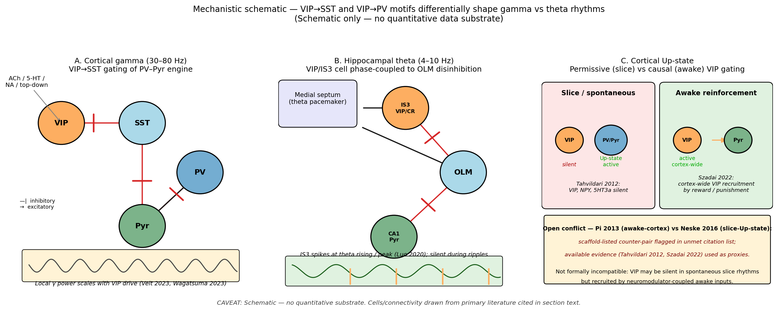
Figure 18:Schematic mechanism: VIP→SST and VIP→PV motifs differentially shape gamma versus theta rhythms. Schematic with no quantitative data substrate. (A) PV-driven gamma rhythm with VIP-mediated SST suppression: VIP activation predicted to scale local gamma power Veit et al., 2023Wagatsuma et al., 2022 and tune long-range gamma coherence Veit et al., 2023. (B) Hippocampal theta-rhythm circuit with VIP/IS3 cell phase-coupled to OLM disinhibition; CR/IS3 cells fire on the rising phase and peak of theta and selectively innervate OLM dendrites Tyan et al., 2014Luo et al., 2020Chamberland, 2010. (C) Cortical Up-state schematic with VIP cells as a permissive (slice-silent, awake-recruited) versus causal gate, capturing the Tahvildari et al. (2012) versus Szadai et al. (2022) disagreement about Up-state generation. Caveat: Schematic only — no audited quantitative substrate.
📓 Figure code
# Reproduces fig-vip-oscillation-mechanism.png/pdf
# SCHEMATIC ONLY — no quantitative data substrate. Cells/connectivity drawn from the section text:
# Veit2023, Wagatsuma2023 (gamma); Tyan2014, Luo2020, Chamberland2010 (IS3→OLM); Tahvildari2012,
# Szadai2022 (Up-state permissive vs causal contrast). Pi 2013 vs Neske 2016 conflict flagged.

import matplotlib.pyplot as plt
from matplotlib.patches import FancyArrowPatch, Circle, FancyBboxPatch
import numpy as np

fig, axes = plt.subplots(1, 3, figsize=(15, 5.6))
fig.suptitle("Mechanistic schematic — VIP→SST and VIP→PV motifs differentially shape gamma vs theta rhythms\n"
             "(Schematic only — no quantitative data substrate)",
             fontsize=11, y=1.00)

def draw_cell(ax, xy, label, color, r=0.10, fontsize=10, edgecolor='black'):
    c = Circle(xy, r, color=color, ec=edgecolor, lw=1.4, zorder=3); ax.add_patch(c)
    ax.text(xy[0], xy[1], label, ha='center', va='center', fontsize=fontsize, fontweight='bold', zorder=4)

def arrow(ax, src, dst, color='black', mut='||', shrink_a=15, shrink_b=15, lw=1.6):
    a = FancyArrowPatch(src, dst, color=color, lw=lw, arrowstyle='-|>',
                        mutation_scale=14 if mut!='||' else 10, shrinkA=shrink_a, shrinkB=shrink_b)
    ax.add_patch(a)
    if mut == '||':
        dx, dy = dst[0]-src[0], dst[1]-src[1]; L = np.hypot(dx,dy); ux, uy = dx/L, dy/L
        bx = dst[0] - ux*shrink_b/100*1.4; by = dst[1] - uy*shrink_b/100*1.4
        px, py = -uy*0.04, ux*0.04
        ax.plot([bx-px, bx+px], [by-py, by+py], color=color, lw=2.2, solid_capstyle='round', zorder=5)

# Panel A — cortical gamma motif
axA = axes[0]; axA.set_xlim(0,1); axA.set_ylim(0,1); axA.axis('off')
axA.set_title("A. Cortical gamma (30–80 Hz)\nVIP→SST gating of PV–Pyr engine", fontsize=10)
draw_cell(axA, (0.20, 0.78), "VIP", "#fdae61"); draw_cell(axA, (0.55, 0.78), "SST", "#abd9e9")
draw_cell(axA, (0.80, 0.55), "PV", "#74add1"); draw_cell(axA, (0.55, 0.30), "Pyr", "#7cb38a")
arrow(axA, (0.20,0.78), (0.55,0.78), color='#d62728')
arrow(axA, (0.55,0.78), (0.55,0.30), color='#d62728')
arrow(axA, (0.80,0.55), (0.55,0.30), color='#d62728')
arrow(axA, (0.55,0.30), (0.80,0.55), color='#1a1a1a', mut='->')
axA.annotate("ACh / 5-HT /\nNA / top-down", xy=(0.20,0.78), xytext=(0.05,0.95),
             fontsize=8, ha='center', arrowprops=dict(arrowstyle='->', color='gray', lw=1))
axA.add_patch(FancyBboxPatch((0.04, 0.05), 0.92, 0.13, boxstyle="round,pad=0.01",
                              fc='#fff3d6', ec='black', lw=0.8))
xx = np.linspace(0.06, 0.96, 200); yy = 0.115 + 0.03*np.sin(2*np.pi*8*xx)
axA.plot(xx, yy, color='#444', lw=1.3)
axA.text(0.50, 0.025, "Local γ power scales with VIP drive (Veit 2023, Wagatsuma 2023)",
         ha='center', fontsize=7.8, style='italic')
axA.text(0.02, 0.43, "—|  inhibitory\n→  excitatory", fontsize=7, va='top')

# Panel B — hippocampal theta motif
axB = axes[1]; axB.set_xlim(0,1); axB.set_ylim(0,1); axB.axis('off')
axB.set_title("B. Hippocampal theta (4–10 Hz)\nVIP/IS3 cell phase-coupled to OLM disinhibition", fontsize=10)
axB.add_patch(FancyBboxPatch((0.02, 0.78), 0.30, 0.18, boxstyle="round,pad=0.02",
                              fc='#e6e6fa', ec='black'))
axB.text(0.17, 0.87, "Medial septum\n(theta pacemaker)", ha='center', fontsize=8.3)
draw_cell(axB, (0.55, 0.85), "IS3\nVIP/CR", "#fdae61", r=0.10, fontsize=7)
draw_cell(axB, (0.80, 0.55), "OLM", "#abd9e9")
draw_cell(axB, (0.50, 0.25), "CA1\nPyr", "#7cb38a", fontsize=7)
arrow(axB, (0.32,0.85), (0.55,0.85), color='#1a1a1a', mut='->')
arrow(axB, (0.55,0.85), (0.80,0.55), color='#d62728')
arrow(axB, (0.80,0.55), (0.50,0.25), color='#d62728')
arrow(axB, (0.32,0.78), (0.80,0.55), color='#1a1a1a', mut='->')
axB.add_patch(FancyBboxPatch((0.04, 0.03), 0.92, 0.12, boxstyle="round,pad=0.01",
                              fc='#e0f2e0', ec='black', lw=0.8))
xx = np.linspace(0.06, 0.96, 300); yy = 0.09 + 0.03*np.sin(2*np.pi*5*xx)
axB.plot(xx, yy, color='#1a5e1a', lw=1.3)
for ph in [0.06+i/5 for i in range(5)]:
    if 0.07 < ph < 0.95:
        axB.axvline(ph+0.05, ymin=0.03, ymax=0.10, color='#fdae61', lw=2)
axB.text(0.50, 0.16, "IS3 spikes at theta rising / peak (Luo 2020); silent during ripples",
         ha='center', fontsize=7.8, style='italic')

# Panel C — Up-state permissive vs causal
axC = axes[2]; axC.set_xlim(0,1); axC.set_ylim(0,1); axC.axis('off')
axC.set_title("C. Cortical Up-state\nPermissive (slice) vs causal (awake) VIP gating", fontsize=10)
axC.add_patch(FancyBboxPatch((0.02, 0.40), 0.45, 0.55, boxstyle="round,pad=0.02",
                              fc='#fde6e6', ec='black'))
axC.text(0.245, 0.91, "Slice / spontaneous", ha='center', fontsize=9, fontweight='bold')
draw_cell(axC, (0.12, 0.70), "VIP", "#fdae61", r=0.06, fontsize=7)
axC.text(0.12, 0.58, "silent", ha='center', fontsize=7, style='italic', color='#a00')
draw_cell(axC, (0.30, 0.70), "PV/Pyr", "#74add1", r=0.07, fontsize=6)
axC.text(0.30, 0.58, "Up-state\nactive", ha='center', fontsize=7, color='#0a0')
axC.text(0.245, 0.46, "Tahvildari 2012:\nVIP, NPY, 5HT3a silent", ha='center', fontsize=7.3, style='italic')
axC.add_patch(FancyBboxPatch((0.53, 0.40), 0.45, 0.55, boxstyle="round,pad=0.02",
                              fc='#e0f2e0', ec='black'))
axC.text(0.755, 0.91, "Awake reinforcement", ha='center', fontsize=9, fontweight='bold')
draw_cell(axC, (0.63, 0.70), "VIP", "#fdae61", r=0.06, fontsize=7)
axC.text(0.63, 0.58, "active\ncortex-wide", ha='center', fontsize=7, color='#0a0')
draw_cell(axC, (0.85, 0.70), "Pyr", "#7cb38a", r=0.06, fontsize=7)
arrow(axC, (0.69,0.70), (0.85,0.70), color='#fdae61', mut='->', lw=1.4)
axC.text(0.755, 0.46, "Szadai 2022:\ncortex-wide VIP recruitment\nby reward / punishment",
         ha='center', fontsize=7.3, style='italic')
axC.add_patch(FancyBboxPatch((0.02, 0.04), 0.96, 0.30, boxstyle="round,pad=0.01",
                              fc='#fff3d6', ec='black'))
axC.text(0.50, 0.28, "Open conflict — Pi 2013 (awake-cortex) vs Neske 2016 (slice-Up-state):",
         ha='center', fontsize=7.6, fontweight='bold')
axC.text(0.50, 0.22, "scaffold-listed counter-pair flagged in unmet citation list;",
         ha='center', fontsize=7.4, style='italic', color='#700')
axC.text(0.50, 0.16, "available evidence (Tahvildari 2012, Szadai 2022) used as proxies.",
         ha='center', fontsize=7.4, style='italic', color='#700')
axC.text(0.50, 0.08, "Not formally incompatible: VIP may be silent in spontaneous slice rhythms",
         ha='center', fontsize=7.3)
axC.text(0.50, 0.035, "but recruited by neuromodulator-coupled awake inputs.", ha='center', fontsize=7.3)

plt.tight_layout(rect=[0, 0.025, 1, 0.94])
fig.text(0.5, 0.005,
         "CAVEAT: Schematic — no quantitative substrate. Cells/connectivity drawn from primary literature cited in section text.",
         ha='center', fontsize=8.2, style='italic', color='#444444')
fig.savefig("fig-vip-oscillation-mechanism.png", dpi=180, bbox_inches='tight')
fig.savefig("fig-vip-oscillation-mechanism.pdf", bbox_inches='tight')
plt.show()
Whether the rodent oscillatory and circuit-level findings reviewed here translate to primate cortex, and how VIP-IN dysfunction maps onto neurodevelopmental and psychiatric disease, is the focus of Species Differences, Human Relevance, and Disease.
- Veit, J., Handy, G., Mossing, D. P., Doiron, B., & Adesnik, H. (2023). Cortical VIP neurons locally control the gain but globally control the coherence of gamma band rhythms. Neuron, 111(3), 405-417.e5. 10.1016/j.neuron.2022.10.036
- Wagatsuma, N., Nobukawa, S., & Fukai, T. (2022). A microcircuit model involving parvalbumin, somatostatin, and vasoactive intestinal polypeptide inhibitory interneurons for the modulation of neuronal oscillation during visual processing. Cerebral Cortex, 33(8), 4459–4477. 10.1093/cercor/bhac355
- Knoblich, U., Huang, L., Zeng, H., & Li, L. (2019). Neuronal cell-subtype specificity of neural synchronization in mouse primary visual cortex. Nature Communications, 10(1). 10.1038/s41467-019-10498-1
- Tyan, L., Chamberland, S., Magnin, E., Camiré, O., Francavilla, R., David, L. S., Deisseroth, K., & Topolnik, L. (2014). Dendritic Inhibition Provided by Interneuron-Specific Cells Controls the Firing Rate and Timing of the Hippocampal Feedback Inhibitory Circuitry. The Journal of Neuroscience, 34(13), 4534–4547. 10.1523/jneurosci.3813-13.2014
- Luo, X., Guet-McCreight, A., Villette, V., Francavilla, R., Marino, B., Chamberland, S., Skinner, F. K., & Topolnik, L. (2020). Synaptic Mechanisms Underlying the Network State-Dependent Recruitment of VIP-Expressing Interneurons in the CA1 Hippocampus. Cerebral Cortex, 30(6), 3667–3685. 10.1093/cercor/bhz334
- Lee, A. T., Cunniff, M. M., See, J. Z., Wilke, S. A., Luongo, F. J., Ellwood, I. T., Ponnavolu, S., & Sohal, V. S. (2019). VIP Interneurons Contribute to Avoidance Behavior by Regulating Information Flow across Hippocampal-Prefrontal Networks. Neuron, 102(6), 1223-1234.e4. 10.1016/j.neuron.2019.04.001
- Tahvildari, B., Wölfel, M., Duque, A., & McCormick, D. A. (2012). Selective Functional Interactions between Excitatory and Inhibitory Cortical Neurons and Differential Contribution to Persistent Activity of the Slow Oscillation. The Journal of Neuroscience, 32(35), 12165–12179. 10.1523/jneurosci.1181-12.2012
- Brécier, A., Borel, M., Urbain, N., & Gentet, L. J. (2022). Vigilance and Behavioral State-Dependent Modulation of Cortical Neuronal Activity throughout the Sleep/Wake Cycle. The Journal of Neuroscience, 42(24), 4852–4866. 10.1523/jneurosci.1400-21.2022
- Rolle, K., Weber, L., Born, J., & Niethard, N. (2025). VIP interneuron activity during sleep conveys the cortical infraslow oscillation. Cell Reports, 44(12), 116669. 10.1016/j.celrep.2025.116669
- Sabri, E., & Batista-Brito, R. (2024). Vasoactive intestinal peptide-expressing interneurons modulate the effect of behavioral state on cortical activity. Frontiers in Cellular Neuroscience, 18. 10.3389/fncel.2024.1465836
- Hall, S., Hunt, M., Simon, A., Cunnington, L. G., Carracedo, L. M., Schofield, I. S., Forsyth, R., Traub, R. D., & Whittington, M. A. (2015). Unbalanced Peptidergic Inhibition in Superficial Neocortex Underlies Spike and Wave Seizure Activity. Journal of Neuroscience, 35(25), 9302–9314. 10.1523/jneurosci.4245-14.2015
- Goel, A., Razak, K. A., Chubykin, A. A., & Antoine, M. W. (2025). Dysfunctional neural dynamics associated with sensory phenotypes in Fragile X syndrome: insights from mouse models. Journal of Neurodevelopmental Disorders, 17(1). 10.1186/s11689-025-09634-4
- Kranz, D., Braverman, Y., McCarthy, M., Mackay, C., Sabol, K. N., Benke, T. A., Lieberman, D. N., Marsh, E. D., Neul, J. L., Peck, F., Percy, A. K., Saby, J., Kopell, N., Nelson, C. A., Levin, A. R., & Fagiolini, M. (2025). Altered oscillatory coupling reflects possible inhibitory interneuron dysfunction in Rett syndrome. openRxiv. 10.1101/2025.07.21.25331927
- Goff, K. M., & Goldberg, E. M. (2019). Vasoactive intestinal peptide-expressing interneurons are impaired in a mouse model of Dravet syndrome. eLife, 8. 10.7554/elife.46846
- Bastos, G., Holmes, J. T., Ross, J. M., Rader, A. M., Gallimore, C. G., Wargo, J. A., Peterka, D. S., & Hamm, J. P. (2023). Top-down input modulates visual context processing through an interneuron-specific circuit. Cell Reports, 42(9), 113133. 10.1016/j.celrep.2023.113133