Cortical VIP interneurons are members of the caudal-ganglionic-eminence (CGE)–derived 5-HT3AR / Adarb2 lineage that also gives rise to the Lamp5, Sncg and Serpinf1 subclasses Lee et al., 2010Miyoshi et al., 2010Tasic et al., 2018. The cells are born in a late embryonic neurogenic wave, migrate tangentially along several caudo-rostral streams, settle preferentially in superficial layers, and acquire mature firing, synaptic and morphological properties through a protracted postnatal programme that combines cell-intrinsic transcription-factor cascades with experience- and activity-dependent refinement Miyoshi et al., 2010Miyoshi et al., 2015Lim et al., 2018De Marco García & Fishell, 2024. Two features of this developmental programme matter most for the rest of this review. First, the gene-regulatory logic that distinguishes VIP from sister CGE classes is now traceable to a small set of transcription factors — most notably Prox1, Sp8/Sp9 and COUP-TFII (Nr2f2) — whose conditional perturbation produces subtype-selective phenotypes Miyoshi et al., 2015Stachniak et al., 2021Wei et al., 2019Kanatani et al., 2008. Second, every quantitative claim about CGE-lineage proportion or VIP-subtype identity that derives from mouse fate-mapping must be re-examined when applied to primate cortex, where lineage proportions, marker codes and progenitor architecture diverge Hodge et al., 2019Hansen et al., 2013Ma et al., 2013Marín, 2025. The remainder of this section reviews each step in turn, and explicitly flags the cross-species comparisons that complicate translation.
CGE origin and the 5-HT3AR / Adarb2 lineage
The mapping of GABAergic subtypes onto subpallial origins is one of the most extensively replicated facts in cortical development. Homochronic transplantation of medial-ganglionic-eminence (MGE) versus caudal-ganglionic-eminence (CGE) explants into host mouse embryos established that PV and SST cardinal classes derive primarily from the MGE, whereas reelin-, calretinin-, NPY- and VIP-expressing cardinal classes derive primarily from the CGE Bandler et al., 2017. Joint contributions of progenitor spatial origin, birthdate, lineage relationships and division mode further refine subtype identity, as resolved at scRNA-seq resolution by Mi et al. (2018). Inducible Mash1/Olig2-CreER fate mapping subsequently quantified the CGE contribution at ~30% of all cortical interneurons across at least nine distinct mature subtypes, with CGE neurogenesis peaking at E16.5 — about three days later than MGE neurogenesis — and 75% of CGE progeny populating supragranular layers regardless of birthdate Miyoshi et al., 2010. A parallel hippocampal study using transgenic and inducible reporter lines documented the same temporal staggering: an MGE wave between E9–E12 generates PV and SST hippocampal interneurons, whereas a CGE wave between E12–E16 generates the CCK+, calretinin+, VIP+ and reelin+ populations that populate stratum lacunosum-moleculare and deep stratum radiatum.
Within this CGE-derived population, the serotonin 5-HT3A receptor (encoded by Htr3a) provides a near-complete molecular handle. Lee et al. (2010) showed that 5-HT3AR expression demarcates essentially all neocortical GABAergic interneurons that lack PV/SST and labels the entire CGE lineage in mouse cortex Lee et al., 2010. Homochronic in utero grafts of E14 Htr3a-GFP+ cells into host cortex reproduce the supragranular laminar bias and the neurochemical profile (VIP, NPY, reelin, calretinin) characteristic of CGE-derived interneurons Vucurovic et al., 2010. Modern scRNA-seq taxonomies recapitulate this division at the transcriptomic level: in mouse cortex, CGE origin maps onto the Vip, Lamp5, Sncg and Serpinf1 subclasses Tasic et al., 2018, while in human middle temporal gyrus the CGE branch is identified by ADARB2 expression and accounts for ~50% of cortical interneurons against ~44% for the LHX6+ MGE branch Hodge et al., 2019. The CGE label therefore unifies a heterogeneous group of upper-layer interneurons whose shared origin is more easily defined than any single mature feature.
Two clarifications are needed. First, the CGE is not the only ventral source of cortical inhibitory cells: the embryonic preoptic area (POA) generates a smaller, mostly reelin+ population that contributes to layer-1 NGC-like cells and that — because POA-derived cells share PROX1, NR2F2 and reelin expression with CGE-derived NGCs — partially confounds CGE-only fate maps Gelman et al., 2009Niquille et al., 2018. Second, the dorsal MGE contributes a calretinin+/SST+ lineage (controlled in part by Nkx6-2) that overlaps in marker space with CGE-derived calretinin+ VIP cells but corresponds to Martinotti-like rather than VIP-bipolar morphologies. These caveats explain why marker-only assignments (e.g. “CR+” or “5-HT3AR+”) give an imperfect read-out of CGE lineage and why fate-mapping with intersectional driver lines remains the gold standard.
Transcription-factor cascades that specify VIP and its CGE-class neighbours
The transcription-factor (TF) logic that specifies CGE-derived interneurons differs in detail from the Lhx6/Sox6-dominated MGE programme Liodis et al., 2007. Single-cell transcriptomic profiling of E13.5 MGE and E14.5 CGE/LGE reveals a conserved mitotic-progenitor maturation trajectory across all three eminences, onto which eminence-specific TF cassettes are overlaid in postmitotic precursors to seed cardinal-class diversity Mayer et al., 2018; chromatin-accessibility profiling shows that enhancer remodelling precedes transcriptional commitment, with CGE-specific Prox1, Sp8 and COUP-TFII programmes opening in coordinated waves Allaway et al., 2021. By P0, scRNA-seq separates a Lhx6-negative postmitotic CGE branch into Vip-, Id2- and Sncg-aligned precursor states already distinct from MGE-derived progeny Mayer et al., 2018.
Among the CGE-class TFs, Prox1 has the strongest causal evidence. PROX1 is expressed in 84% of fate-mapped CGE/LGE/POA-derived interneurons and in 100% of VIP+ cortical interneurons in P30 mouse cortex Rubin & Kessaris, 2013, and is selectively maintained in postmitotic CGE-derived interneuron precursors as the first identified CGE-specific postmitotic TF Miyoshi et al., 2015. Embryonic conditional deletion of Prox1 impairs the integration of CGE-derived precursors into superficial layers and differentially regulates the postnatal maturation of RELN+, Calb2/VIP+ and VIP+ subtypes — establishing PROX1 as the first identified TF specifically required for CGE-lineage acquisition of subtype-specific properties. Postmitotic Prox1 ablation in early postnatal VIP cells then reveals an additional, subtype-selective role: it reduces Elfn1-dependent short-term plasticity of excitatory inputs onto multipolar but not bipolar VIP cells, in line with adult VIP-subclass transcriptomic preservation of Prox1 expression Tasic et al., 2018Hodge et al., 2019.
COUP-TFII (NR2F2) is preferentially expressed in CGE relative to MGE and is required for the caudal migratory stream that channels CGE progeny into caudal cortex, hippocampus and amygdala Kanatani et al., 2008. In adult mouse cortex, Cai2013 showed that COUP-TFII is also expressed in dorsal MGE-, dorsal LGE- and POA-derived cells, so COUP-TFII positivity is not a clean CGE-only marker Cai et al., 2012. In hippocampus, COUP-TFII is selectively maintained in CGE-derived calretinin+, nNOS+, reelin+, neurogliaform and CCK/calbindin+ basket cells Fuentealba et al., 2010. Conditional COUP-TFI inactivation perturbs both lateral and medial CGE migratory streams and disrupts SP8/COUP-TFII expression, producing laminar-specific deficits across distinct interneuron subtypes in adult cortex Touzot et al., 2016. Human relevance: NR2F1 (COUP-TFI) loss-of-function causes Bosch–Boonstra–Schaaf optic atrophy syndrome (intellectual disability, autism, seizures) Yang et al., 2017, and COUP-TFII is broadly expressed in human fetal forebrain GE and neocortex from 9–22 gestational weeks with peak in the CGE Reinchisi et al., 2011.
Sp8 and Sp9 define a second axis of CGE/dLGE specification. Sp8 is expressed in ~20% of adult cortical interneurons derived from both dorsal LGE and dorsal CGE, weakly in MGE mantle but strongly in CGE/dLGE Tao et al., 2019, and acts cell-autonomously to specify olfactory-bulb and cortical CGE-related lineages. Co-expressed Sp8/Sp9 in the dorsal CGE SVZ jointly drive CGE-derived interneuron specification in part by repressing Pak3, Robo1 and Slit1 and by activating Cxcl14 Wei et al., 2019, while Sp9 alone is required in MGE for normal PV/SST development Liu et al., 2018. The mutually exclusive deployment of Sp9 in MGE versus Sp8/Sp9 in CGE thus partly explains the cardinal-class divergence captured in adult taxonomies. Lhx6 itself is required not for GABAergic identity but for MGE-specific specification and migration; in Lhx6−/− mice some MGE cells aberrantly acquire a CGE-like identity, and in vivo complementation shows that Arx and Cxcr7 rescue separable aspects of Lhx6 function Alifragis et al., 2004Zhao et al., 2008.
Several additional regulators affect VIP-relevant subsets. Dlx1/Dlx2 and Dlx5/Dlx6 control CGE neurite outgrowth and tangential migration, and selective Dlx1 loss causes subtype-specific reduction of CR+, SST+ and other cortical/hippocampal interneurons Cobos et al., 2007Wang et al., 2010Cobos et al., 2005. CXCL12 attracts immature interneurons during tangential migration via CXCR4/CXCR7 streams in subpial and intermediate-zone routes, and BDNF/NT4 acting via TrkB stimulates tangential migration of MGE-derived cells, with a residual effect on CGE-associated calbindin+ populations. The net picture, reviewed by Lim et al. (2018) and De Marco García & Fishell (2024), is that cardinal-class identity (including VIP) is largely seeded by progenitor-stage TF combinations, while finer t-type granularity emerges through postmitotic refinement that integrates intrinsic TF cascades with activity-dependent signalling.
Tangential migration and laminar settling
CGE-derived precursors leave the eminence in three caudo-rostral streams whose temporal sequence and TF coding differ from the dorsoventral MGE streams. Initial studies Touzot et al., 2016 resolved a lateral migratory stream (LMS), a medial migratory stream (MMS) and the previously known caudal migratory stream (CMS), each expressing distinct combinations of SP8, PROX1, COUP-TFI and COUP-TFII; conditional COUP-TFI inactivation differentially perturbs LMS/MMS and produces laminar-specific deficits in adult cortex. Lopezbendito2004 used GAD65-GFP transgenic mice to track a CGE-enriched subpopulation along the lower intermediate zone from E14–E15 and into superficial layers thereafter, consistent with the late-born CGE programme. Foundational studies Bellion, 2005 characterised the cellular biomechanics of tangential migration as a two-phase nucleokinesis (centrosome–Golgi forward translocation followed by nuclear advance) — a mode shared between MGE- and CGE-derived precursors. The chemokine CXCL12 acting via CXCR4/CXCR7 attracts cortical interneurons during these tangential streams, with loss of CXCR4 responsiveness producing dispersion deficits, and BDNF/TrkB signalling provides additional permissive drive that affects both MGE- and CGE-associated populations.
Laminar settling is initially comparable between MGE and CGE: at P1, mouse precursors born at E12.5 from either eminence show similar layer distributions, with subsequent radial sorting between deep (MGE) and superficial (CGE) layers driven by mechanisms that coincide with KCC2 upregulation Miyoshi & Fishell, 2010. By the end of the first postnatal week, CGE-derived cells have completed their characteristic preferential occupancy of layers II/III (~75%) Miyoshi et al., 2010, although in mouse this enrichment is graded rather than absolute and a substantial CGE-derived contingent settles in deeper layers and in layer 1 Lee et al., 2010. Larimer2016 used heterochronic CGE transplants into neonatal mouse V1 to show that CGE-derived precursors disperse, acquire host-like laminar position, marker expression, intrinsic properties and visual response — but, unlike MGE transplants, they do not reactivate ocular dominance plasticity, with the residual ODP induction observed in CGE transplants attributable to a small contaminating fraction of MGE-lineage cells.
A subtle but consequential observation is that excitatory-neuron identity instructs lineage-specific aspects of CGE settling. Conditional Satb2 knockout in mouse projection neurons reprograms intratelencephalic-type pyramidal neurons toward a pyramidal-tract identity and selectively disrupts the lamination and circuit integration of CGE-derived (but not MGE-derived) interneurons, providing causal evidence that pyramidal subtype identity instructs CGE-lineage interneuron development in a lineage-specific manner. Cux1 and Cux2 are themselves expressed cell-autonomously in postmitotic Reelin+ cortical interneurons — a Reelin+ population that includes both MGE-derived neurogliaform and CGE-derived components — and are required for the development of this Reelin-secreting IN subset Cubelos et al., 2008, placing Cut-family homeodomain TF control inside the same superficial-layer compartment that CGE-class lateral and medial migratory streams populate Touzot et al., 2016. The chemoattractant Cxcl14, induced by Sp8/Sp9, similarly couples CGE migration choices to layer-specific cues during the same window. This intercellular dependency complicates simple cell-autonomous accounts of VIP laminar settling and dovetails with the activity-dependent programmes considered next.
Activity-dependent and intrinsic programmes for postnatal maturation
Postnatal maturation of CGE-derived interneurons depends on a graded interplay between cell-intrinsic genetic programmes and activity-dependent extrinsic signals. The seminal cell-autonomous experiment is that of De Marco García et al. (2011), who showed by in utero Dlx5/6-eGFP electroporation at E15.5 followed by Kir2.1-mediated silencing that activity is essential before P3 for correct migration of CGE-derived reelin+ and calretinin+ (but not VIP+) interneurons; after P3, glutamate-mediated activity controls axon and dendrite development. The Dlx1 target Elmo1 mediates this activity-dependent migration in Re+/Cr+ subtypes and is downregulated by Kir2.1 expression De Marco García et al., 2011. Established work extended these findings using the same Kir2.1 paradigm, reporting that cell-autonomous activity reduction lowered maximum AP discharge from 93±10 Hz to 44±8 Hz and broadened AP half-width from 1.3±0.15 ms to 3.0±0.32 ms in CGE-derived cells (P12–P15 cortex), and produced subtype-specific laminar mispositioning in Re+ and Cr+ but not VIP+ cells Karayannis et al., 2012. The convergence of these two studies established the disinhibitory view that CGE interneuron migration and intrinsic excitability mature through cell-autonomous activity, with VIP cells comparatively spared.
Glutamatergic drive is required for the survival and synaptic maturation of CGE-derived interneurons more broadly. Conditional removal of all AMPAR subunits (GluA1/2/3 cKO) in CGE-derived interneurons increases their early postnatal numbers but causes later apoptosis to converge with wild-type, almost completely eliminates sEPSCs, and reduces feedforward and feedback inhibition onto pyramidal neurons. The phenotype indicates that AMPAR-mediated excitatory drive is required not for CGE-IN birth but for circuit integration and survival during the postnatal critical period. Within VIP cells specifically, Simacek et al. (2025) showed that inhibitory presynaptic inputs onto layer 2/3 VIP-INs in mouse barrel cortex undergo functional maturation between P9 and P15: both readily-releasable vesicle number and release probability rise to adult-like values across this window. The complementary excitatory side is captured by Earlier work Simacek et al., 2025, which described a sequential P3 → P30+ pattern in mouse S1 barrel cortex in which mEPSC frequency rises before P8–10, intrinsic firing matures next, and IPSC kinetics consolidate last. The Prox1 / Elfn1 axis additionally specifies short-term excitatory input dynamics in a VIP-subtype-selective manner during the same window Stachniak et al., 2021, and this maturation timeline is anchored to the broader CGE-class postnatal programme described in recent reviews Lim et al., 2018De Marco García & Fishell, 2024.
Two reviews integrate these strands. Lim et al. (2018) argued that cortical GABAergic interneuron diversity emerges from cell-intrinsic genetic programmes in subpallial progenitors that unfold over a protracted period and are subsequently modulated by activity-dependent, non-cell-autonomous mechanisms during circuit integration; De Marco García & Fishell (2024) updated this synthesis with new Patch-seq and connectivity data, emphasising that transient developmental connectivity instructs the consolidation of mature subtypes. Yaeger et al. (2019) provided a circuit-level example in mouse V1, where basal-forebrain acetylcholine directly excites SST interneurons during the critical period to enable compartmentalised dendritic spiking; this cholinergic responsiveness is lost in adulthood, and suppressing SST cells during the critical period prevents normal development of binocular receptive fields. While VIP cells were not the primary focus, the experiment illustrates the principle that experience-dependent neuromodulator drive and CGE-class circuit function are functionally coupled during postnatal critical periods, with implications for VIP-driven disinhibitory motifs that emerge in the same window.
Programmed cell death, network activity, and circuit integration
Roughly a third of cortical interneurons die during the second postnatal week in a developmental wave of programmed cell death (PCD). Previous reports showed that all ventral-eminence-derived cortical interneurons — including CGE-derived cells — undergo PCD, but that VIP interneuron survival, in contrast to MGE-derived PV/SST classes, is not activity-dependent; calcineurin in interneurons sequentially controls morphological maturation (E15–P5) and subsequent activity-dependent processes Priya et al., 2018. This decoupling matters: it means that cell-autonomous reductions in VIP excitability (as in De Marco García et al. (2011)) do not preferentially cull VIP cells, whereas comparable manipulations bias the survival of CGE reelin+/calretinin+ and MGE-derived populations. Carriere et al. (2020) provided a parallel cell-autonomous mechanism: clustered γ-Protocadherins (Pcdhgs) act in mouse GABAergic interneurons to promote survival during the postnatal PCD critical period, with effects across both MGE- and CGE-derived classes.
Network-level activity feeds back onto interneuron survival. Duan et al. (2020) reported that in neonatal mouse somatosensory cortex (P7), functional interneuron and pyramidal-cell assemblies emerge spatially segregated; reducing GABA release or synaptic inputs onto pyramidal cells erodes this functional topography and increases interneuron survival, demonstrating that GABAergic restriction of early network dynamics actively regulates interneuron developmental death. Together with the AMPAR-cKO data Akgül et al., 2019, the Pcdhg result Carriere et al., 2020, and the activity-independence of VIP-cell survival reported by Priya et al. (2018), these findings recast the second postnatal week as a coordinated period in which intrinsic survival modules and circuit-level activity are layered onto the earlier, lineage-specific transcriptional cascades — with VIP cells representing a partial exception whose survival is set by intrinsic rather than activity-driven programmes De Marco García & Fishell, 2024.
A handful of studies extend this developmental account to specific VIP-relevant circuits. In hippocampal CA1, a subset of VIP+ interneurons projects long-range axons to subiculum and is recruited preferentially during quiet wakefulness rather than during theta-run/locomotion, indicating that sublineage diversification of CA1 VIP cells produces functionally specialised long-range projection cells distinct from the disinhibitory VIP-IS3 population Francavilla et al., 2018. The hippocampal CGE neurogenic wave that produces these populations is itself temporally protracted from E12 to E16, generating not only IS3-type VIP cells but also nNOS+ NGC and Ivy populations from a shared CGE/MGE-mixed origin. In V1, Ibrahim et al. (2021) showed that bottom-up thalamic inputs to layer-1 NGCs are required postnatally to establish higher-order top-down connectivity onto these cells, sculpting circuit maturation in a CGE-class-relevant compartment. Cortically deployed VIP cells thus mature into multiple functionally distinct sublineages whose relative proportions vary by area and species — a point that becomes acute when the analysis crosses to primates.
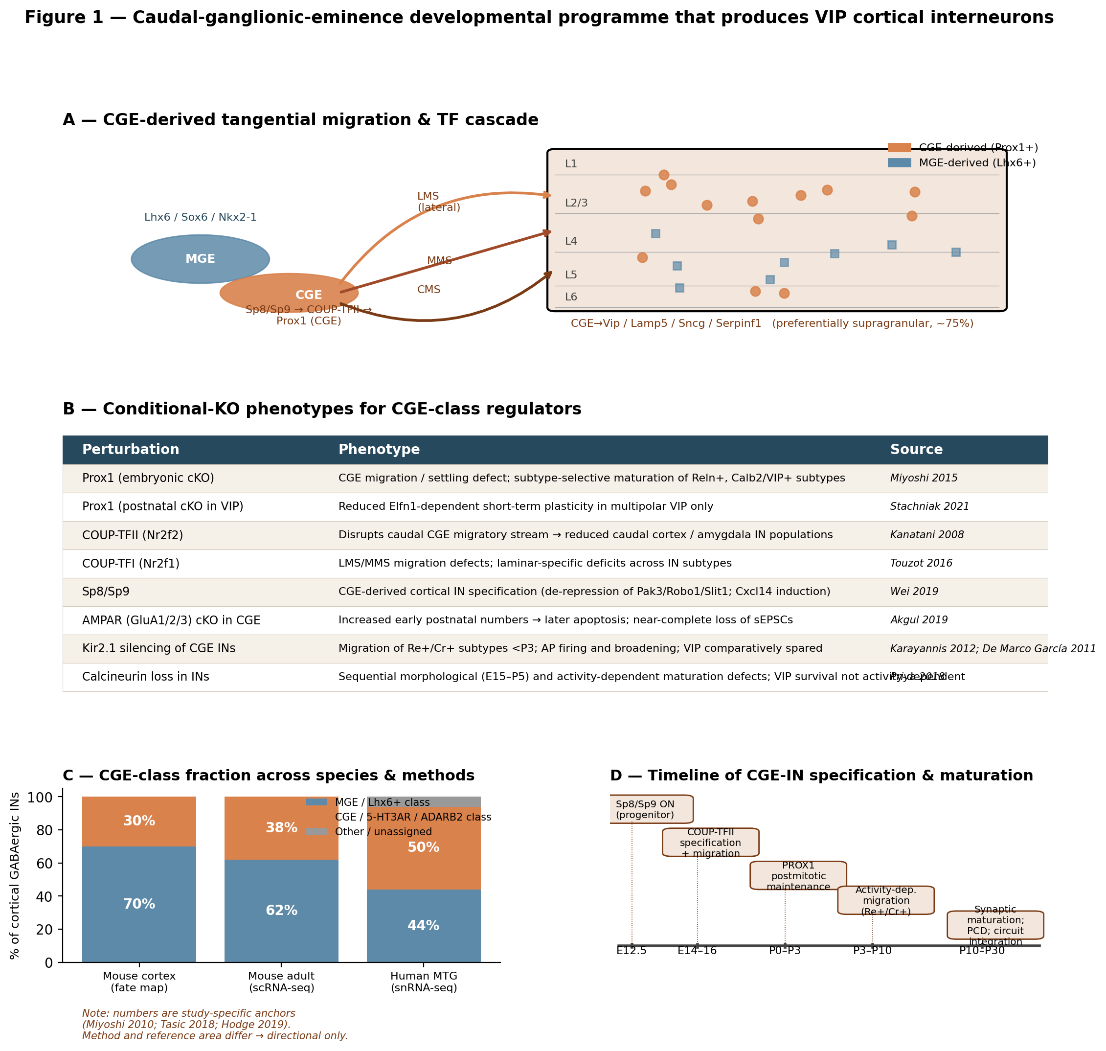
Figure 3:Schematic of the CGE-derived inhibitory lineage that produces VIP interneurons. (A) Embryonic CGE → tangential migration via lateral, medial and caudal streams → cortical layer settling. CGE-class TFs (Prox1, Sp8, Nr2f2/COUP-TFII) are contrasted with the MGE-class TF Lhx6 Miyoshi et al., 2010Touzot et al., 2016Kanatani et al., 2008Liodis et al., 2007. (B) Conditional-KO phenotypes for the principal CGE TFs and synaptic/excitability regulators, showing the subtype-selectivity of each perturbation Miyoshi et al., 2015Stachniak et al., 2021Cai et al., 2012Touzot et al., 2016Wei et al., 2019Akgül et al., 2019Karayannis et al., 2012De Marco García et al., 2011Priya et al., 2018. (C) Reported fraction of cortical GABAergic neurons attributable to the CGE/5-HT3AR/ADARB2 lineage in mouse fate-mapping (~30%) versus human snRNA-seq (~50%) Miyoshi et al., 2010Hodge et al., 2019. Caveat: the cross-species comparison overlays a fate-map percentage (mouse) against a marker-defined snRNA-seq percentage (human MTG only), so the apparent expansion conflates measurement modality with biology and should be interpreted as a directional rather than absolute estimate.
📓 Figure code
import matplotlib.pyplot as plt
import matplotlib.patches as mpatches
import numpy as np

# Panel A: schematic of MGE/CGE → migration streams → cortex laminar pattern.
# Panel B: TF-cKO phenotype table (Prox1, COUP-TFI/II, Sp8/Sp9, AMPAR, Kir2.1, Calcineurin).
# Panel C: cross-species CGE-class fraction (30% mouse fate-map / ~38% mouse Tasic 2018 / 50% human Hodge 2019).
# Panel D: TF cascade timeline (Sp8/Sp9 → COUP-TFII → PROX1 → activity-dep. → synaptic maturation).

# Anchor values used in Panel C:
anchors = {
    'Mouse cortex (fate map, Miyoshi 2010)':   {'CGE': 30, 'MGE': 70, 'Other': 0},
    'Mouse adult (scRNA-seq, Tasic 2018)':     {'CGE': 38, 'MGE': 62, 'Other': 0},
    'Human MTG (snRNA-seq, Hodge 2019)':       {'CGE': 50, 'MGE': 44, 'Other': 6},
}
for k,v in anchors.items():
    print(k, v)

print('Schematic — see fig-cge-lineage.png/.pdf for rendered output.')
Cross-species divergence: rodent fate-mapping versus primate transcriptomics
The transition from rodent to primate complicates almost every quantitative claim made above. Ma et al. (2013) argued from immunohistochemistry of Sox6, COUP-TFII and Sp8 in developing human and monkey telencephalon that the same three subpallial progenitor domains (MGE, LGE, CGE) found in rodents are present in primates and that tangentially migrating interneurons retain comparable marker codes — supporting a conserved subcortical origin of primate neocortical interneurons Ma et al., 2013. Marín (2025) synthesised the broader comparative literature and concluded that interneuron diversity is largely conserved between rodents and primates, with primate cortical evolution contributing greater abundance and wider interconnectivity rather than wholly new types Marín, 2025. Single-cell taxonomies are consistent: comparative scRNA-seq across macaques and mice indicates that prenatal IN classes are largely conserved, even if a primate-specific TAC3 striatal interneuron population emerges Schmitz et al., 2022, and snRNA-seq of >2.4 million cells across 18 marmoset locations shows that adult transcriptomic identity tracks developmental origin more strongly than neurotransmitter repertoire Krienen et al., 2023.
Conserved cardinality, however, masks several quantitative and architectural divergences. Earlier studies Hansen et al., 2013 documented a massively expanded subventricular zone in second-trimester human ganglionic eminences that lacks the radial-fibre/oriented organisation seen in rodents and contains stem cells whose proliferation and migration patterns differ from the MGE programme — implying human-specific aspects of CGE-lineage development. Prior work Reinchisi et al., 2011 confirmed that COUP-TFII expression peaks in human CGE during gestational weeks 9–22 with a temporo-occipital > frontal/dorsal gradient. scRNA-seq of human ganglionic eminences identifies regional and temporal progenitor diversity and an evolutionarily conserved transcriptional grammar but also human-specific transcriptomic features, while Earlier reports Velmeshev et al., 2023 profiled >700,000 nuclei across 106 prenatal/postnatal human cortex donors and prioritised interneuron lineages as key vulnerable trajectories. Eichmüller et al. (2022) further described a human-specific caudal late interneuron progenitor (CLIP) population that over-proliferates in tuberous sclerosis complex organoids — a primate-relevant lineage component absent in mouse fate maps.
The most consequential primate divergence for VIP biology is in lineage marker codes. Hodge et al. (2019) reported that human MTG contains roughly equal proportions of MGE-derived (44% LHX6+) and CGE-derived (50% ADARB2+) cortical interneurons, that VIP is the most transcriptomically diverse interneuron subclass in human MTG (21 types), and — crucially — that the disinhibitory mouse CGE marker HTR3A is not expressed in human CGE-lineage interneuron types, marking a major species-specific divergence in the very molecular handle that defined the CGE lineage in mouse Hodge et al., 2019. The same study identified human-specific features within the CGE class (e.g., some CGE-class layer-1 cells co-express SST, a feature not seen in mouse layer-1 interneurons) and located human “rosehip” cells transcriptomically within the CGE-class LAMP5 subdivision Hodge et al., 2019. Initial studies Jorstad et al., 2023 confirmed across eight human cortical areas that 24 cell subclasses are highly consistent in presence but show areal differences in inhibitory-neuron sub-state composition.
A further methodological note: the disinhibitory adult mouse interneuron taxonomy of Tasic et al. (2018) defines six GABAergic subclasses (Sst, Pvalb, Vip, Lamp5, Sncg, Serpinf1) plus two distinct types — placing VIP as one of four CGE-class subclasses. Reported VIP fractions vary across studies and atlas integrations because Lamp5, Sncg and Serpinf1 may be pooled with VIP under a “5-HT3AR class” or split out depending on clustering resolution; reconciling proportions therefore requires that subclass boundaries be specified, and recent cross-species human/marmoset comparisons Hodge et al., 2019Krienen et al., 2023 reinforce that subclass-resolution choices have first-order consequences for any cross-paper percentage. The Velthoven et al. (2025) developmental atlas resolves these subclass boundaries at prenatal-to-postnatal resolution and provides a hierarchical anchor for cross-study reconciliation. Beyond cortex, Sorrells et al. (2019) showed that the human amygdala paralaminar nuclei develop adjacent to the CGE and contain immature DCX+/PSA-NCAM+ neurons that mostly express excitatory markers and transition to mature TBR1+/VGLUT2+ neurons during adolescence — establishing a protracted, CGE-adjacent developmental programme in human amygdala distinct from the cortical CGE-IN lineage that other comparative studies emphasise.
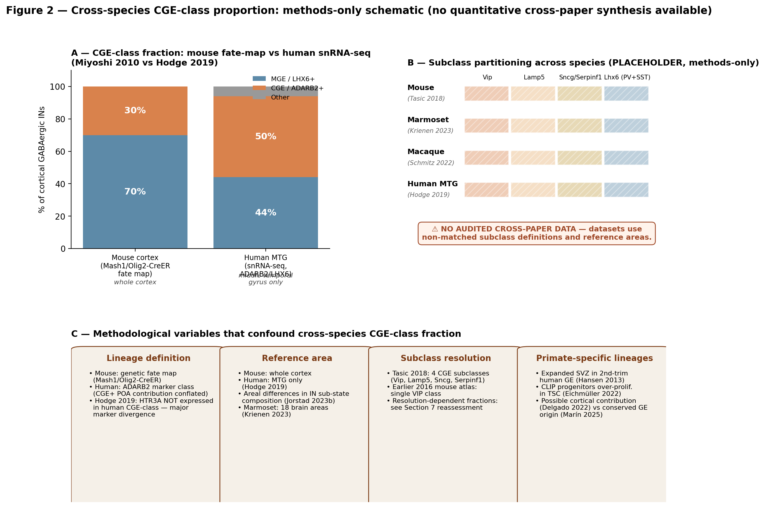
Figure 4:Schematic methods-only summary of the cross-species CGE-lineage comparison. (A) Reported fraction of cortical GABAergic neurons assigned to the CGE/5-HT3AR/ADARB2 class — mouse fate-mapping (Mash1/Olig2-CreER) gives ~30% across whole cortex Miyoshi et al., 2010; human snRNA-seq in MTG gives ~50% (LHX6+ MGE 44% / ADARB2+ CGE 50%) Hodge et al., 2019. (B) The proposed partitioning across VIP / Lamp5 / Sncg / Serpinf1 subclasses across mouse, marmoset, macaque and human is shown as a methods placeholder only: no audited cross-paper panel was achievable for this stacked comparison because the underlying datasets use non-matched subclass definitions and reference areas Tasic et al., 2018Hodge et al., 2019Krienen et al., 2023Schmitz et al., 2022. (C) Side timeline of the CGE-progenitor TF cascade — Sp8/Sp9 → COUP-TFII → PROX1 → postmitotic Prox1 / Elfn1 / activity-dependent refinement — with conditional-KO outcome annotations Wei et al., 2019Kanatani et al., 2008De Marco García et al., 2011Karayannis et al., 2012. Caveat: No quantitative cross-paper comparison was achievable for this comparison; the figure is methods-only/schematic, with the directional claim (mouse < primate CGE-class fraction) supported but the precise gap dependent on measurement modality. Readers should treat percentage estimates as study-specific anchors rather than as a synthesised effect size.
📓 Figure code
import matplotlib.pyplot as plt
import numpy as np
import matplotlib.patches as mpatches

# Anchor values used in Panel A:
panel_a = {
    'Mouse cortex (Miyoshi 2010, fate map)': {'CGE': 30, 'MGE': 70, 'Other': 0},
    'Human MTG (Hodge 2019, snRNA-seq)':     {'CGE': 50, 'MGE': 44, 'Other': 6},
}
for k,v in panel_a.items():
    print(k, v)

# Panel B is a deliberate placeholder — non-matched subclass definitions across Tasic 2018, Krienen 2023, Schmitz 2022, Hodge 2019.
print('Methods-only schematic — see fig-cge-fraction-cross-species.png/.pdf.')
CGE internal topography, organoid models, and recent integration
The CGE is not internally homogeneous. Foundational studies Garg et al., 2025 showed that mature CGE-IN subtypes arise from distinct spatial and temporal subdomains of the CGE — dorsal/ventral and rostral/caudal subregions and birthdate windows prefigure adult identities, mirroring the internal topography long appreciated in the MGE. Established work Bright et al., 2025 complemented this with heterochronic transplantation, lineage tracing and perturb-seq experiments showing that progenitor competence is reset across embryonic time across all GEs, so that progenitor-stage temporal coding interacts with eminence-of-origin to set cardinal identity. Bandler et al. (2017) had previously argued that cortical interneuron specification is governed by the joint contributions of spatial origin, birthdate, lineage relationships and mode of cell division — a framework that the more recent CGE-resolved data Garg et al., 2025Bright et al., 2025 now operationalise. The 2025 review of Horton & Paredes (2025) synthesises CGE-derived IN biology across mouse, human and non-human primate, providing the most current cross-species integration. Velthoven et al. (2025) adds a prenatal–postnatal scRNA-seq dataset of >1.2 million mouse telencephalon cells (E7–P14) that resolves the hierarchical GABAergic taxonomy at developmental resolution and anchors the prenatal-to-postnatal trajectory of CGE-class precursors.
Stem-cell and organoid systems are increasingly used to model CGE-lineage development, with mixed fidelity. Bershteyn et al. (2023) defined the directed-differentiation cascade (NKX2-1+/SOX1+/FOXG1+/DLX1/2+ → MGE progenitors → mature interneurons) for pallial MGE-type cells from human ESCs; analogous CGE protocols, exemplified by Earlier work Du et al., 2025, generate ~80% calretinin+ cells via SHH activation plus IWP2-mediated WNT inhibition that show GABAergic action potentials in vitro and integrate after transplantation. Previous reports Andrews et al., 2023 showed that dorsal forebrain organoids initially generate LGE-like INs at week 8 and progressively replace them with CGE-like INs by week 15, with LIF/LIFR signalling promoting outer radial glia identity in primary tissue. Uzquiano et al. (2022) profiled human cortical organoids at single-cell, epigenetic and spatial levels and reported that endogenous cellular diversification trajectories are recapitulated regardless of metabolic state. Eichmüller et al. (2022)’s identification of a human-specific CLIP cell population in TSC organoids underscores that organoid systems are now uncovering primate-specific CGE-class progenitor states absent in mouse. The translational corollary is that mouse fate-mapping should anchor the framework but human-specific organoid and snRNA-seq data must be consulted for any quantitative claim about VIP-class proportions or marker codes in primate cortex.
A specific point about cortical NGCs and amygdala / ventral telencephalon connections is worth preserving here. Tang et al. (2012) showed that COUP-TFII is required in the CGE to specify amygdala (BMA) patterning by directly activating semaphorin receptors Nrp1 and Nrp2; in conditional COUP-TFII mutants, CGE-derived migration to amygdala is disrupted, indicating that CGE-class TFs serve patterning functions beyond cortex. Mercier et al. (2022) complements the cortical story by showing that hippocampal stratum-lacunosum-moleculare interneurons (including NGCs) exhibit NMDAR-dependent Hebbian LTP, with plasticity in NGC inputs modulating temporoammonic excitation-inhibition balance — providing a circuit-level rationale for the developmental investment in CGE-class hippocampal NGCs documented by Tricoire et al. (2010) and Tricoire et al. (2011). Kim et al. (2025) reported that neonatal gyrencephalic brains (human, non-human primate, piglet) harbour an expanded curved subventricular zone (‘Arc’) of migratory neuroblasts absent in lissencephalic mouse and marmoset, adding yet another lineage-relevant primate-specific feature that mouse fate-mapping cannot capture.
A short note on hippocampal CA1 VIP development. The hippocampal CGE wave (E12–E16) generates the disinhibitory IS3-type VIP/CR/CCK CA1 interneuron alongside CCK+ basket and NGC populations Tricoire et al., 2011Fuentealba et al., 2010. CA1 VIP cells diversify postnatally into at least two distinct subpopulations: the local IS3-type disinhibitory cells described in early reviews Rudy et al., 2010Kepecs & Fishell, 2014 and the long-range VIP-LRP cells with subicular projections that preferentially fire during quiet wakefulness rather than during theta-run/locomotion Francavilla et al., 2018. The latter population breaks the disinhibitory assumption that all CA1 VIP cells implement the same disinhibitory motif and indicates that within-VIP sublineage diversification — likely set by the same Prox1/Sp8/COUP-TFII cascade documented for cortex but acting on hippocampus-specific targets — produces functionally distinct outputs that (Local Circuit Motifs and the Disinhibition Framework) will revisit.
Synthesis: what the development data buy the rest of the review
The developmental literature delivers four claims that the rest of this review will lean on. (1) VIP cortical interneurons are CGE-derived members of a 5-HT3AR/ADARB2-defined sister set (with Lamp5, Sncg, Serpinf1) that is reproducible across modalities and species at the cardinal-class level Lee et al., 2010Miyoshi et al., 2010Tasic et al., 2018Hodge et al., 2019. (2) Their identity is specified by a small, conditional-KO-validated TF cascade (Sp8/Sp9 → COUP-TFII → PROX1 → postmitotic PROX1 / Elfn1) whose perturbation produces subtype-selective cellular and synaptic phenotypes traceable into adult VIP-IN function Wei et al., 2019Kanatani et al., 2008Miyoshi et al., 2015Stachniak et al., 2021. (3) Their postnatal maturation combines a cell-intrinsic genetic programme with activity-dependent and circuit-level inputs, with VIP cells comparatively spared from the activity-dependent migration and survival mechanisms that affect Re+/Cr+ subtypes De Marco García et al., 2011Karayannis et al., 2012Priya et al., 2018Akgül et al., 2019Lim et al., 2018De Marco García & Fishell, 2024. (4) The translation of mouse fate-mapping to human VIP biology is bounded: lineage marker codes diverge (loss of HTR3A as a CGE handle in human; expansion of CGE-class proportion to ~50% in MTG; appearance of human-specific CLIP and rosehip-type lineages), and quantitative cross-species claims should be made with explicit modality and area caveats Hodge et al., 2019Hansen et al., 2013Reinchisi et al., 2011Eichmüller et al., 2022Marín, 2025. (Morphological Diversity) takes these molecular and developmental categories and asks how they project into morphological space; (Species Differences, Human Relevance, and Disease) returns to the cross-species question for disease-relevant VIP phenotypes; and the synthesis in Local Circuit Motifs and the Disinhibition Framework returns to the resolution-dependence of mouse VIP-fraction estimates flagged above.
- Lee, S., Hjerling-Leffler, J., Zagha, E., Fishell, G., & Rudy, B. (2010). The Largest Group of Superficial Neocortical GABAergic Interneurons Expresses Ionotropic Serotonin Receptors. The Journal of Neuroscience, 30(50), 16796–16808. 10.1523/jneurosci.1869-10.2010
- Miyoshi, G., Hjerling-Leffler, J., Karayannis, T., Sousa, V. H., Butt, S. J. B., Battiste, J., Johnson, J. E., Machold, R. P., & Fishell, G. (2010). Genetic Fate Mapping Reveals That the Caudal Ganglionic Eminence Produces a Large and Diverse Population of Superficial Cortical Interneurons. The Journal of Neuroscience, 30(5), 1582–1594. 10.1523/jneurosci.4515-09.2010
- Tasic, B., Yao, Z., Graybuck, L. T., Smith, K. A., Nguyen, T. N., Bertagnolli, D., Goldy, J., Garren, E., Economo, M. N., Viswanathan, S., Penn, O., Bakken, T., Menon, V., Miller, J., Fong, O., Hirokawa, K. E., Lathia, K., Rimorin, C., Tieu, M., … Zeng, H. (2018). Shared and distinct transcriptomic cell types across neocortical areas. Nature, 563(7729), 72–78. 10.1038/s41586-018-0654-5
- Miyoshi, G., Young, A., Petros, T., Karayannis, T., McKenzie Chang, M., Lavado, A., Iwano, T., Nakajima, M., Taniguchi, H., Huang, Z. J., Heintz, N., Oliver, G., Matsuzaki, F., Machold, R. P., & Fishell, G. (2015). Prox1Regulates the Subtype-Specific Development of Caudal Ganglionic Eminence-Derived GABAergic Cortical Interneurons. The Journal of Neuroscience, 35(37), 12869–12889. 10.1523/jneurosci.1164-15.2015
- Lim, L., Mi, D., Llorca, A., & Marín, O. (2018). Development and Functional Diversification of Cortical Interneurons. Neuron, 100(2), 294–313. 10.1016/j.neuron.2018.10.009
- De Marco García, N. V., & Fishell, G. (2024). Interneuron Diversity: How Form Becomes Function. Cold Spring Harbor Perspectives in Biology, 17(7), a041513. 10.1101/cshperspect.a041513
- Stachniak, T. J., Kastli, R., Hanley, O., Argunsah, A. Ö., van der Valk, E. G. T., Kanatouris, G., & Karayannis, T. (2021). Postmitotic Prox1 Expression Controls the Final Specification of Cortical VIP Interneuron Subtypes. The Journal of Neuroscience, 41(39), 8150–8162. 10.1523/jneurosci.1021-21.2021
- Wei, S., Du, H., Li, Z., Tao, G., Xu, Z., Song, X., Shang, Z., Su, Z., Chen, H., Wen, Y., Liu, G., You, Y., Zhang, Z., & Yang, Z. (2019). Transcription factors Sp8 and Sp9 regulate the development of caudal ganglionic eminence-derived cortical interneurons. Journal of Comparative Neurology, 527(17), 2860–2874. 10.1002/cne.24712
- Kanatani, S., Yozu, M., Tabata, H., & Nakajima, K. (2008). COUP-TFII Is Preferentially Expressed in the Caudal Ganglionic Eminence and Is Involved in the Caudal Migratory Stream. The Journal of Neuroscience, 28(50), 13582–13591. 10.1523/jneurosci.2132-08.2008
- Hodge, R. D., Bakken, T. E., Miller, J. A., Smith, K. A., Barkan, E. R., Graybuck, L. T., Close, J. L., Long, B., Johansen, N., Penn, O., Yao, Z., Eggermont, J., Höllt, T., Levi, B. P., Shehata, S. I., Aevermann, B., Beller, A., Bertagnolli, D., Brouner, K., … Lein, E. S. (2019). Conserved cell types with divergent features in human versus mouse cortex. Nature, 573(7772), 61–68. 10.1038/s41586-019-1506-7
- Hansen, D. V., Lui, J. H., Flandin, P., Yoshikawa, K., Rubenstein, J. L., Alvarez-Buylla, A., & Kriegstein, A. R. (2013). Non-epithelial stem cells and cortical interneuron production in the human ganglionic eminences. Nature Neuroscience, 16(11), 1576–1587. 10.1038/nn.3541
- Ma, T., Wang, C., Wang, L., Zhou, X., Tian, M., Zhang, Q., Zhang, Y., Li, J., Liu, Z., Cai, Y., Liu, F., You, Y., Chen, C., Campbell, K., Song, H., Ma, L., Rubenstein, J. L., & Yang, Z. (2013). Subcortical origins of human and monkey neocortical interneurons. Nature Neuroscience, 16(11), 1588–1597. 10.1038/nn.3536
- Marín, O. (2025). Development of GABAergic Interneurons in the Human Cerebral Cortex. European Journal of Neuroscience, 61(9). 10.1111/ejn.70136
- Bandler, R. C., Mayer, C., & Fishell, G. (2017). Cortical interneuron specification: the juncture of genes, time and geometry. Current Opinion in Neurobiology, 42, 17–24. 10.1016/j.conb.2016.10.003
- Mi, D., Li, Z., Lim, L., Li, M., Moissidis, M., Yang, Y., Gao, T., Hu, T. X., Pratt, T., Price, D. J., Sestan, N., & Marín, O. (2018). Early emergence of cortical interneuron diversity in the mouse embryo. Science, 360(6384), 81–85. 10.1126/science.aar6821