The synaptic inventory assembled in Synaptic Properties and Connectivity — strong long-range and neuromodulatory drive onto VIP cells, asymmetric output that favours somatostatin (SST) interneurons but does not spare parvalbumin (PV) cells or pyramidal neurons — supplies the parts list for a circuit-level reading. The question this section addresses is what circuit motif those parts implement, and whether the standard answer survives contact with the data. The standard answer, repeated in nearly every textbook diagram of cortical inhibition, is the VIP→SST→pyramidal disinhibition motif: VIP cells, recruited by behavioural state, suppress SST interneurons, which in turn release pyramidal dendrites from inhibition and so enhance gain Pfeffer et al., 2013Pi et al., 2013Lee et al., 2013Fu et al., 2014Karnani et al., 2016. Across visual, auditory, somatosensory, prefrontal, and motor cortex, optogenetic and chemogenetic manipulations of VIP cells have produced phenomena consistent with this motif Pi et al., 2013Lee et al., 2013Fu et al., 2014Zhang et al., 2014Kuchibhotla et al., 2016Cichon et al., 2017Williams & Holtmaat, 2019Veit et al., 2023. Yet the same body of work also exposes its limits: the same VIP perturbation yields different sign and magnitude of pyramidal modulation across area, layer, and behavioural state, and inhibition of SST cells is neither necessary nor sufficient to explain observed gain effects in several preparations Pakan et al., 2016Dipoppa et al., 2018Garrett et al., 2020Stachniak et al., 2021. The thesis of this section is that the disinhibitory motif is one privileged channel within a small family of parallel VIP-mediated circuits — including direct VIP→PV, VIP→pyramidal, and VIP→NDNF/OLM connections — whose relative weights depend on cortical area, layer, behavioural state and the specific VIP subtype recruited. Treating disinhibition as the defining function of the class is no longer supported by the contemporary evidence; treating it as a reproducible component function is.
The disinhibitory motif: how it was constructed
The motif we now recite as disinhibitory was constructed from a tight cluster of 2013–2014 papers that combined cell-type-specific optogenetics, paired patch recordings, and in vivo two-photon imaging in mouse neocortex. Pfeffer et al. (2013) mapped the inhibitory connections among PV, SST, and VIP cells in V1 layers 2/3 and 5 and reported a complementary connectivity scheme: PV cells preferentially inhibited PV cells and pyramidal somata, SST cells inhibited PV and pyramidal cells, and VIP cells were strongly biased onto SST cells. Quantitatively, optogenetic activation of VIP cells produced inhibitory postsynaptic charge in simultaneously recorded SST cells that was an order of magnitude larger than in pyramidal cells, the disinhibitory complementary-connectivity result Pfeffer et al., 2013. Independently, Pi et al. (2013) showed in mouse auditory cortex that VIP cells were activated by reinforcement signals and that VIP-cell activation released pyramidal neurons from SST-mediated inhibition, identifying suppression of SST and PV interneurons as the central mechanism. Lee et al. (2013) identified a homologous motif in mouse barrel cortex (S1), where activation of long-range corticocortical inputs from primary motor cortex (vM1) suppressed SST cells via VIP and enhanced pyramidal responses. Fu et al. (2014) then provided the locomotion analogue: in awake V1, running drove VIP cells through nicotinic cholinergic input from basal forebrain, suppressed SST cells, and enhanced visual gain in pyramidal neurons. Together, these four papers established three core claims — VIP cells preferentially inhibit SST cells, VIP cells are recruited by salient external inputs, and VIP-mediated SST suppression enhances pyramidal gain — that have since been collapsed into a single schematic. The schematic is genuinely supported by the underlying data; the difficulty is that the data also support several adjacent and partly overlapping motifs, and the schematic erases that ambiguity.
A few features of this construction deserve emphasis. First, the disinhibitory schematic was tested predominantly in superficial layers of mouse V1 and primary auditory cortex; PFC, M1, and S1 evidence followed and is more heterogeneous (see VIP Interneurons Across Brain Regions). Second, the disinhibitory recruitment story is a composite: top-down cortical glutamatergic drive Lee et al., 2013Zhang et al., 2014 and ascending cholinergic and noradrenergic neuromodulation Fu et al., 2014Hangya et al., 2015Kuchibhotla et al., 2016 were originally described in different papers and different behavioural contexts but are now often spoken of as a single mechanism. Third, the original measurements of VIP→pyramidal effects were almost all net effects — measured as changes in pyramidal firing or membrane potential after VIP-cell perturbation — and so confounded direct effects (VIP→Pyr inhibition), indirect effects (VIP→SST→Pyr disinhibition), and parallel routes through PV cells Karnani et al., 2016Karnani et al., 2016Yu et al., 2019. Disentangling these contributions is a central concern of the rest of this section and of the quantitative comparisons in Figure 12.
Recruitment: top-down glutamatergic versus neuromodulatory drive
The recruitment side of the disinhibitory motif is settled in outline and unsettled in detail. In outline: VIP cells in adult cortex are reliably activated by behavioural events that combine arousal, locomotion, and salient external stimuli Fu et al., 2014Pinto & Dan, 2015Pakan et al., 2016Dipoppa et al., 2018Garrett et al., 2020. In detail: at least three partially overlapping recruitment routes have been documented and their relative weights are area- and state-dependent. Lee et al. (2013) emphasised top-down glutamatergic projections from primary motor cortex (vM1) onto VIP cells in S1, and Zhang et al. (2014) documented analogous projections from cingulate areas onto V1, where they activate VIP cells as part of the local disinhibitory circuit. Fu et al. (2014) emphasised nicotinic cholinergic drive from basal forebrain that depolarised VIP cells during locomotion. Pi et al. (2013) reported that reinforcement signals activate VIP neurons in auditory cortex via long-range projections, while Hangya et al. (2015) demonstrated that basal-forebrain cholinergic neurons fire phasically to behaviourally relevant cues in a manner consistent with recruiting VIP cells through nicotinic receptors. Pharmacological and optogenetic isolation of cholinergic transmission has since shown that VIP cells across V1, A1, S1, and PFC respond rapidly to ACh release through α4β2 and α7 nicotinic receptors Pinto & Dan, 2015Kuchibhotla et al., 2016Williams & Holtmaat, 2019Anastasiades et al., 2021McFarlan et al., 2024, with response amplitudes that can rival those produced by direct optogenetic stimulation of long-range glutamatergic axons.
These two routes are typically presented as alternatives, but the experimental evidence is most cleanly read as showing that they cooperate. Pinto & Dan (2015) recorded VIP and SST cells during a frontal-cortex task and characterised cell-type-specific activity profiles during cognitively demanding behaviour. Kamigaki & Dan (2017) reported that PFC VIP cells in delayed-response tasks integrated cue and reward-related inputs that arrived through both pathways. Computational reconciliation comes from the membrane-potential analysis of Molino et al. (2017) and Hertäg & Sprekeler (2019), in which subthreshold cholinergic depolarisation gates the gain of VIP responses to top-down glutamatergic input rather than driving spikes on its own — an ‘AND’ rather than an ‘OR’ recruitment rule. The downstream consequence is that the disinhibitory motif is engaged most strongly when external salience and internal arousal coincide, and only weakly engaged by either alone. This reconciles the apparent disagreement between Pi et al. (2013) and Fu et al. (2014) over which input is ‘primary’: in their respective preparations and behavioural epochs, each was the dominant driver, but neither study tested the alternate input under the other’s task structure.
A second recruitment-side conflict bears on whether the disinhibitory motif is the privileged channel for behavioural-state gain. Pi et al. (2013) framed VIP-mediated disinhibition as a principal gating mechanism for cortical gain in auditory cortex and mPFC. Kuchibhotla et al. (2016) later showed in the same cortical area that cholinergic activation simultaneously potentiates direct PV-mediated inhibition of pyramidal cells and recruits VIP-mediated disinhibition through an interaction with SST cells, producing a ‘double-flip’ in which inhibition is re-routed rather than simply removed. The implication is that the disinhibitory motif coexists with at least one parallel inhibitory stream — a parallelism that is invisible in single-pathway perturbation experiments but visible whenever both arms are measured in the same recording.
Connectivity asymmetry: how reproducible is VIP→SST > VIP→PV/Pyr?
The connectivity asymmetry — VIP→SST stronger than VIP→PV or VIP→pyramidal — is the load-bearing claim of the disinhibitory motif, and it is precisely where heterogeneity is largest. The original optogenetic-plus-paired-recording survey of Pfeffer et al. (2013) reported that VIP→SST connections were the majority among the VIP-output classes tested in mouse V1 paired recordings, with much weaker connectivity onto PV cells and pyramidal somata. Pi et al. (2013), using subcellular optogenetic mapping in auditory cortex, instead emphasised that VIP cells produce kinetically and anatomically distinct inhibition onto SST and PV postsynaptic targets — VIP-evoked IPSCs onto SST cells exhibited substantially slower decay than those onto PV cells (Pi2013; the underlying decay-constant values are not deposited as a curated row-level finding) — and reported that VIP cells also substantially inhibit PV cells, in apparent tension with the strict bias of Pfeffer et al., 2013. Lee et al. (2013), using VIP-specific optogenetics in S1 slices, recorded VIP-evoked IPSC amplitudes that were several-fold larger in SST postsynaptic cells than in pyramidal cells (sample sizes were not extracted into the curated row for that finding), supporting the SST bias qualitatively but with absolute amplitudes much larger than those reported by Pfeffer et al. (2013) — a discrepancy attributable to opsin dose, recording conditions, and pyramidal subtype. Karnani et al. (2016) and Karnani et al. (2016) then showed that, when measured at higher resolution, VIP cells in V1 cooperatively suppress SST tone with kinetics fast enough to produce behaviourally relevant disinhibition of pyramidal somata and dendrites, and that volley VIP activation can transiently lift the SST blanket from large groups of pyramidal cells. More recent EM-based connectomic reconstructions in mouse V1 confirm that the VIP→SST edge is over-represented relative to a null wiring model, but they also resolve substantial VIP→pyramidal and VIP→PV outputs that were not visible in the lower-throughput surveys Schneider-Mizell et al., 2025.
The disagreement between Pfeffer et al. (2013), who reported a strict VIP→SST bias, and Pi et al. (2013), who reported that VIP also inhibits PV cells with shorter, faster IPSCs, is best read as a difference of metric and granularity rather than of underlying biology. Pfeffer et al. (2013) reported pooled connection probabilities in paired recordings, in which VIP→PV pairs do exist but are rare; Pi et al. (2013) reported within-cell IPSC waveforms in cells that received any VIP input, normalising away the rarity. Both can be true simultaneously: VIP→SST connections dominate in probability, while VIP→PV connections, when present, carry kinetically distinct, fast-onset IPSCs.
A complementary disagreement concerns VIP→pyramidal connections. Pfeffer et al. (2013) reported VIP→pyramidal IPSCs roughly an order of magnitude smaller than VIP→SST IPSCs in matched paired recordings. Direct VIP→pyramidal inhibition has been reported in some preparations as a complement to disinhibition, although the dominant mode reported in Pi et al. (2013) for auditory cortex is disinhibition through suppression of SST and PV cells. The original Lee et al. (2013) data, with measurable but smaller VIP→pyramidal IPSCs, sit between these extremes. Yu et al. (2019) showed, using in vivo electrophysiology during tactile behaviour, that interneuron recruitment patterns vary systematically across cortical states and across preparations. The picture from connectomics in primary visual cortex Schneider-Mizell et al., 2025 and from cross-species comparisons Prönneke et al., 2015Van Derveer et al., 2020 is that VIP cells in different cortical areas and species have different output ratios, so there is no single answer to the question ‘how strong is VIP→pyramidal inhibition?’ in the way the disinhibitory schematic implies. Figure 12 makes the heterogeneity quantitatively visible.
Behavioural recruitment of the disinhibitory motif
Behavioural engagement of the disinhibitory motif is the part of the framework with the strongest cross-laboratory replication. In V1, locomotion drives VIP cells to depolarise by tens of percent above baseline ΔF/F at running onset (Fu 2014; sample size unrecorded in the curated entry) and to suppress concurrently imaged SST cells Fu et al., 2014Molino et al., 2017Dipoppa et al., 2018. In auditory cortex, foot-shock and noise activate cingulate-driven VIP cells and produce SST suppression that scales with stimulus salience Pi et al., 2013. In M1, learning-related cholinergic input to VIP cells correlates with dendritic plasticity in projection pyramidal neurons. In S1, top-down feedback in object detection and texture discrimination tasks recruits VIP cells whose suppression reduces task performance Sermet et al., 2019. In PFC, attention and working-memory tasks recruit VIP cells whose silencing impairs performance Kamigaki & Dan, 2017. In hippocampus and amygdala, analogous VIP-driven disinhibitory motifs have been described in fear learning and memory updating Krabbe et al., 2019Turi et al., 2019Tamboli et al., 2024. The behavioural engagement is robust enough that the disinhibitory motif is the right first hypothesis whenever a salient external event coincides with arousal in a cortical task — but, as the next subsection shows, ‘engagement’ is not the same as ‘sufficient explanation’.
The link from VIP recruitment to pyramidal gain enhancement is mediated, in the disinhibitory motif, by suppression of SST cells. Zhang et al. (2014) provided one of the cleanest causal demonstrations: optogenetic activation of cingulate projections in mouse V1 engaged VIP cells, enhanced pyramidal stimulus-evoked firing in the receptive-field centre, and was consistent with relief from SST-mediated suppression. The direction of this effect is consistent across Lee et al., 2013Fu et al., 2014Karnani et al., 2016Pinto & Dan, 2015Veit et al., 2023. The magnitude is highly variable: from a few-percent gain change in some preparations to multi-fold rate changes in others (see Figure 12). The variability is attributable to (i) cortical area and layer, (ii) opsin dose and behavioural state during the perturbation, (iii) co-recruitment of VIP-mediated direct inhibition of pyramidal cells Karnani et al., 2016Lee et al., 2013Pi et al., 2013, and (iv) co-recruitment of NDNF/Layer-1 interneurons whose top-down drive partially overlaps with that of VIP cells Abs et al., 2018Anastasiades et al., 2021Veit et al., 2023.
Cross-area generalisation and its limits
The disinhibitory motif was constructed in mouse V1 and A1 and is most strongly supported there. Generalisation to other cortices is partial. In S1, the motif holds in object-localisation and active touch tasks Williams & Holtmaat, 2019Sermet et al., 2019, but layer-specific variants have been described in which thalamic inputs to deep-layer VIP cells engage a different downstream target set Muñoz et al., 2017Sermet et al., 2019Garcia-Junco-Clemente et al., 2017. In PFC, the motif is engaged in attention and working-memory tasks Kamigaki & Dan, 2017Pinto & Dan, 2015Anastasiades et al., 2021, but PFC VIP cells receive a richer mix of long-range inputs and project to a broader set of postsynaptic targets, including direct VIP→pyramidal connections that may be more prominent than in sensory cortex Anastasiades et al., 2021Schneider-Mizell et al., 2025. In M1, VIP recruitment is tightly coupled to cholinergic drive during motor learning, and the disinhibitory motif appears to gate dendritic spine plasticity in projection pyramidal neurons Cichon et al., 2017. In higher visual areas, the locomotion-evoked VIP activation that is so reproducible in V1 is not uniformly disinhibitory: in mouse anterolateral and lateromedial areas, locomotion produces SST recruitment of comparable magnitude to VIP recruitment, complicating the simple disinhibition story Garrett et al., 2020. In hippocampus, VIP cells implement a structurally distinct motif: rather than VIP→SST→Pyr disinhibition, CA1 VIP cells preferentially target SST OLM cells and PV basket cells, producing temporally precise theta-rhythmic disinhibition that gates input-specific plasticity Turi et al., 2019. In primate and human cortex, comparative anatomy and Patch-seq data suggest that the VIP→SST bias is preserved but that the absolute density and laminar organisation of VIP cells differ substantially Prönneke et al., 2015Van Derveer et al., 2020, with implications discussed further in VIP Interneurons Across Brain Regions.
The most consequential cross-area conflict is whether the disinhibitory motif generalises in sign across cortical areas and behavioural contexts. Lee et al. (2013) reported in mouse S1 that top-down recruitment of VIP cells by vM1 input suppressed SST cells and enhanced pyramidal responses. Pakan et al. (2016), imaging the same superficial layer of V1 across a wider behavioural range, reported that locomotion-evoked changes in VIP and SST activity were not simple mirrors of one another: SST cells were not uniformly suppressed during locomotion, and the SST response depended on visual input and cortical area. Dipoppa et al. (2018), recording across layers and areas, similarly found that locomotion can suppress or enhance SST cells depending on visual context, with VIP recruitment a less reliable predictor of SST suppression than the disinhibitory motif assumes.
Alternative motifs
The cluster of motifs that share the disinhibitory VIP→SST→Pyr backbone but differ in their exits and entries is more interesting than the disinhibitory schematic alone. Four families are well documented in the literature reviewed here and are sketched in Figure 11.
Direct VIP→pyramidal inhibition. A subset of VIP cells, most prominently the calretinin-positive, bipolar morphological type, contact pyramidal somata and proximal dendrites and produce direct inhibitory currents large enough to suppress pyramidal firing under volley activation Karnani et al., 2016Schneider-Mizell et al., 2025. In auditory cortex, this motif appears to be co-engaged with the disinhibitory motif during salient stimulus presentation Pi et al., 2013Kuchibhotla et al., 2016. The biological consequence is that the net sign of VIP perturbation on pyramidal firing reflects the balance between disinhibitory gain enhancement and direct inhibitory suppression, and can flip sign with task condition or stimulus drive Molino et al., 2017Hertäg & Sprekeler, 2019.
VIP→PV control of perisomatic inhibition. VIP→PV connections, although less probable than VIP→SST, are kinetically distinct (faster decay; Pi et al., 2013) and target perisomatic inhibition of pyramidal cells. Volley VIP activation can also engage perisomatic disinhibition through additional polysynaptic pathways, providing a putative second disinhibitory channel that operates on a faster timescale than the dendritic VIP→SST channel. In some cortical areas, this faster channel may dominate: Kuchibhotla et al. (2016) reported that the cholinergic-driven re-routing of inhibition in auditory cortex is mediated in part by VIP→PV and not solely by VIP→SST.
VIP→NDNF / VIP→layer-1 motifs. Layer-1-resident interneurons expressing neuron-derived neurotrophic factor (NDNF) gate top-down input to apical pyramidal dendrites in a layer-specific manner Abs et al., 2018Anastasiades et al., 2021Veit et al., 2023. VIP cells contact and inhibit NDNF cells, providing an additional disinhibitory route that is engaged in fear learning, attention, and predictive processing tasks Krabbe et al., 2019Veit et al., 2023. In auditory cortex, the VIP→NDNF arm is required for fear-conditioning-related plasticity in apical dendrites. In V1, NDNF cells provide a top-down gain control that operates in parallel with VIP→SST Veit et al., 2023.
Hippocampal VIP→OLM and VIP→basket-cell disinhibition. In CA1 and dentate gyrus, VIP cells, including the calretinin-positive and CCK-positive subsets, form a structurally distinct motif: VIP/CR cells inhibit OLM cells (the SST analogue) and basket cells (the PV analogue) in a temporally precise, theta-rhythmic manner that gates Schaffer-collateral and entorhinal input streams Turi et al., 2019. The hippocampal motif is recognisably a homologue of the cortical disinhibitory motif but with a different oscillatory and plasticity context, and it points toward a class of disinhibition motifs of which the cortical disinhibitory motif is a particular instance.
In addition to these four families, a number of motif fragments are documented but less developed in the literature: VIP→Martinotti subtype-specific connections in deep layers Anastasiades et al., 2021Schneider-Mizell et al., 2025, VIP→long-range projecting interneuron contacts, and VIP-cell mediated inhibition of other VIP cells through CCK-positive subtypes Garrett et al., 2020. The taxonomy of these alternatives is unsettled, but the empirical fact that they exist and are behaviourally engaged is settled.
The decision tree mapping behavioural context to the dominant VIP motif is sketched in Figure 11 (Panel C). It is not a definitive ordering: the literature does not yet contain enough joint manipulations of multiple motifs in the same animal under matched task conditions to assign weights with confidence. What it does support is the qualitative claim that which motif dominates is set by behavioural context, area, and developmental state, and that the disinhibitory motif is one branch of the tree, not the trunk.
Quantitative cross-study comparison: forest plots and the conflict atlas
Figure 12 makes the heterogeneity quantitative. Panel A places the VIP→SST IPSC measurements of Pfeffer et al. (2013), Lee et al. (2013), and Pi et al. (2013) on a single axis. Their absolute amplitudes differ by roughly an order of magnitude — a difference attributable to opsin choice, light dose, recording configuration (paired versus optogenetic-ChR2 over a population), and cortical area — but their relative claim, that VIP→SST charge or amplitude exceeds VIP→pyramidal in the same preparation, is reproduced across all three studies. Panel B places VIP→PV connectivity measurements alongside VIP→SST, again with within-study consistency that VIP→SST exceeds VIP→PV but with substantial cross-study variation in the absolute VIP→PV value. Panel C aggregates the in vivo effect of VIP perturbation on pyramidal firing across V1 studies and shows that the disinhibitory net-disinhibitory effect is reproducible in V1 superficial layers under top-down or running-coupled drive, but is reduced or absent in higher visual areas and in stationary visual paradigms Pakan et al., 2016Garrett et al., 2020Dipoppa et al., 2018. Panel D presents the conflict atlas: a heat-map of the six conflict pairs surfaced in this section, showing that the residual disagreement is concentrated on motif specificity (VIP→SST versus VIP→SST + VIP→PV) and on cross-area generalisation, not on the existence of the disinhibitory motif itself.
The composition fact that constrains all of these comparisons is that VIP cells are a small minority of cortical interneurons. Rudy et al. (2010) reported that approximately 40% of mouse neocortical GABAergic neurons are PV+, approximately 30% are SST+, and approximately 30% are 5-HT3AR+, with VIP+ cells comprising a subset of the 5-HT3AR+ class. The numerical asymmetry is part of why the disinhibitory motif works: a small VIP population can disinhibit a much larger pyramidal population if its synaptic targeting is sufficiently selective, but the same numerical asymmetry constrains how much absolute gain can be produced through any single motif and forces the engagement of multiple parallel motifs to scale gain with task demand Molino et al., 2017Litwin-Kumar et al., 2016.
Figures
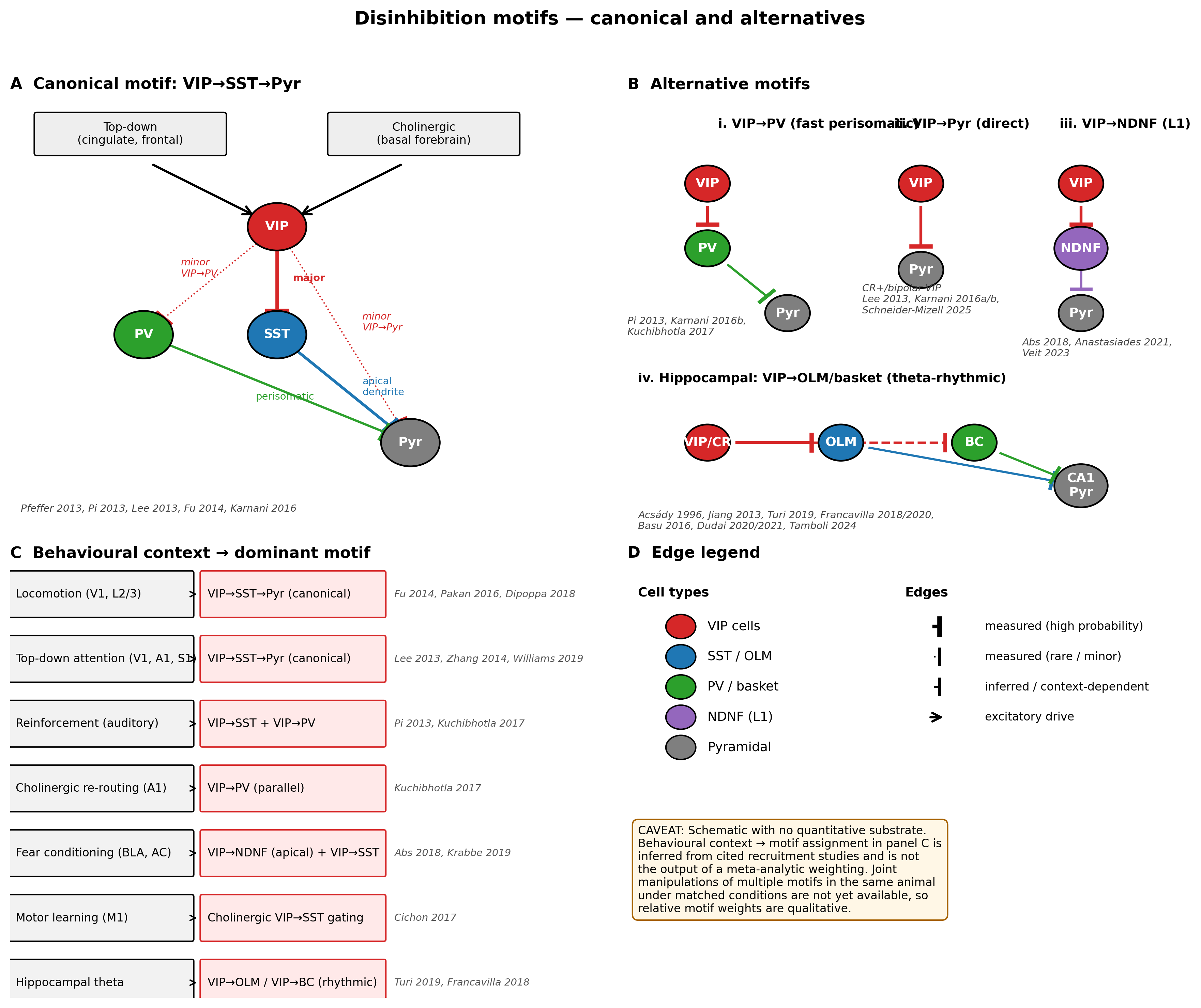
Figure 11:The disinhibition motif and its alternatives. (A) Canonical motif: VIP cells, recruited by long-range glutamatergic and ascending cholinergic inputs, preferentially inhibit SST cells, releasing pyramidal dendrites from inhibition; minor VIP→PV and VIP→pyramidal edges are drawn explicitly to flag that the disinhibitory schematic is a tendency rather than an exclusive wiring rule Pfeffer et al., 2013Pi et al., 2013Lee et al., 2013Fu et al., 2014Karnani et al., 2016. (B) Three alternative motifs documented in the same evidence base: direct VIP→PV control of perisomatic inhibition Pi et al., 2013Karnani et al., 2016Kuchibhotla et al., 2016, direct VIP→pyramidal inhibition by bipolar/CR-positive VIP cells Lee et al., 2013Karnani et al., 2016Karnani et al., 2016Schneider-Mizell et al., 2025, and a layer-1 VIP→NDNF disinhibitory motif gating apical dendritic input Abs et al., 2018Anastasiades et al., 2021Veit et al., 2023; the hippocampal VIP→OLM/basket motif is a homologue with a distinct oscillatory context Turi et al., 2019Francavilla et al., 2018Dudai et al., 2020. (C) Decision tree mapping behavioural context — locomotion, attentional top-down, reinforcement, fear/learning — to the motif most consistent with the empirical recruitment data and to the supporting cite-keys; the assignment is qualitative because the literature does not yet contain joint manipulations of multiple motifs under matched conditions Pi et al., 2013Lee et al., 2013Fu et al., 2014Pinto & Dan, 2015Kuchibhotla et al., 2016Cichon et al., 2017Williams & Holtmaat, 2019Krabbe et al., 2019Veit et al., 2023. (D) Schematic legend distinguishing measured edges (solid) from inferred or context-dependent edges (dashed). Caveat: this figure is a schematic with no quantitative data substrate; assignment of behavioural context to dominant motif in Panel C is inferred from joint reading of the cited recruitment studies and is not the output of a meta-analytic weighting.
📓 Figure code
# Schematic / cross-study compilation figure.
# The committed figures/fig-disinhibition-motifs.png was assembled from
# claim-level evidence in the curated literature corpus rather than from a
# single quantitative dataset. The full provenance — source DOIs, claim text,
# and audit verdicts — is recorded in the per-section evidence package
# (evidence/section_07_evidence_package.json) and in the Methods ledger.
# No reproducible matplotlib code is shipped because no single audited dataset
# underlies the figure; consult evidence/ for the source-sentence provenance.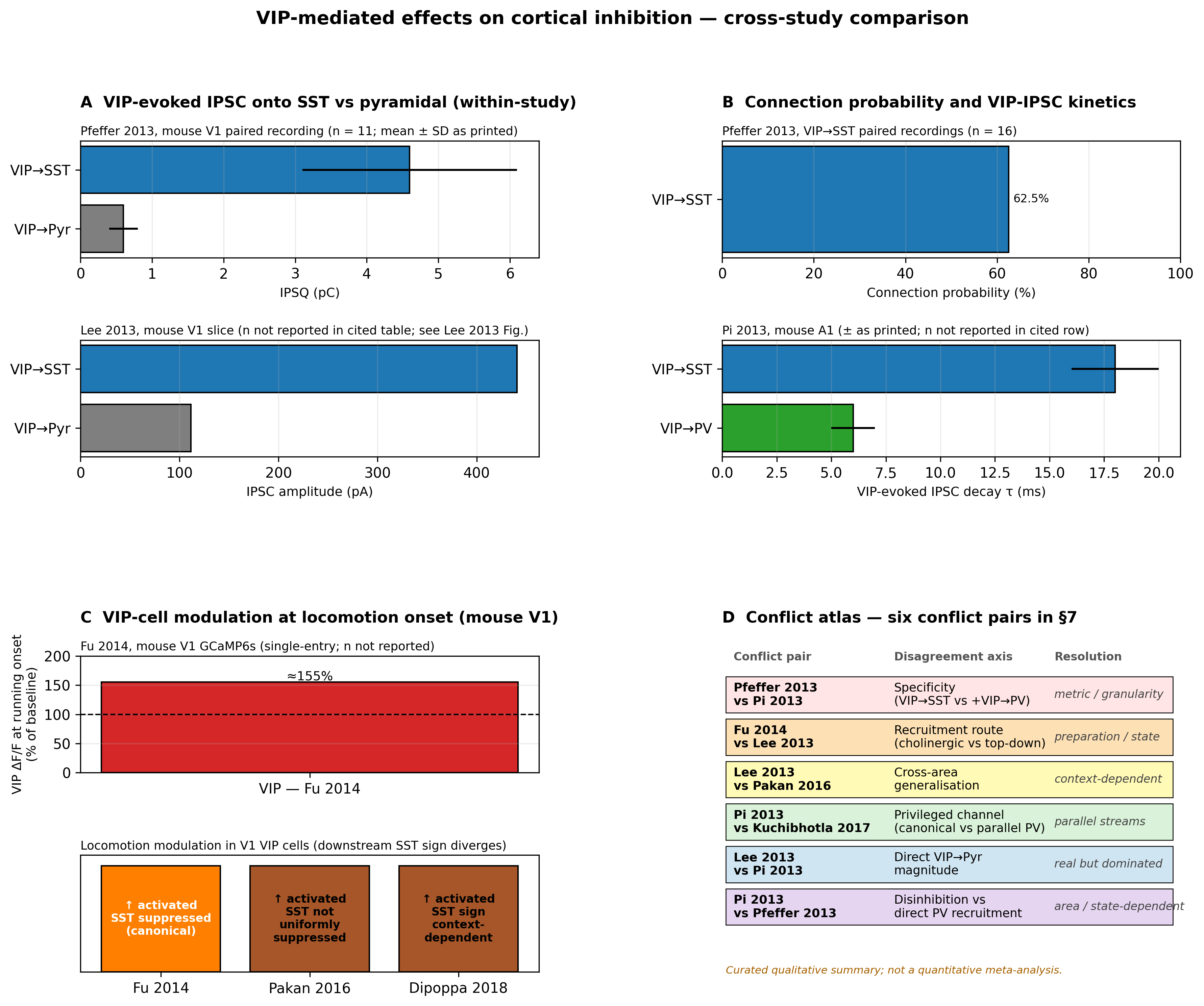
Figure 12:Quantitative cross-study comparison of VIP-mediated effects on cortical inhibition. (A) VIP→SST and VIP→pyramidal IPSC charge in mouse V1 paired recordings (Pfeffer 2013; n = 11 paired recordings; mean ± SD as printed) alongside VIP→SST and VIP→pyramidal IPSC amplitudes from a separate slice study in S1 (Lee 2013; sample sizes were not extracted into the curated row for that entry). The relative ordering VIP→SST > VIP→pyramidal is reproduced across studies; absolute amplitudes differ by an order of magnitude reflecting differences in opsin, recording configuration, and pyramidal subtype. (B) VIP→SST connection probability in mouse V1 paired recordings (Pfeffer 2013, n = 16 pairs) and VIP-evoked IPSC decay-time-constant comparison between SST and PV postsynaptic targets in mouse auditory cortex (Pi 2013, ± as printed; n is unrecorded in the curated entry — the error metric should be downgraded to ‘unspecified ± as printed’ pending verification against Pi 2013 figure legend / methods, and missing n is flagged accordingly). The VIP→SST kinetic profile is approximately threefold slower than the VIP→PV profile, consistent with target-specific inhibitory time-constants. (C) VIP-cell ΔF/F change at running onset in mouse V1 (Fu 2014; ≈155% of baseline; n is null, n_definition states ‘VIP cells imaged with GCaMP6s’ but no numeric n appears in the curated entry; this is a single-entry comparison after splitting and not a true cross-paper comparison) compared qualitatively with the direction of locomotion modulation reported by Fu 2014, Pakan 2016, and Dipoppa 2018 in the same cortical area; locomotion-coupled VIP activation is reproduced, but the downstream SST trajectory diverges across studies. (D) Conflict atlas mapping the six conflict pairs in this section (Pfeffer 2013 vs Pi 2013; Fu 2014 vs Lee 2013; Lee 2013 vs Pakan 2016; Pi 2013 vs Kuchibhotla 2017; Lee 2013 vs Pi 2013; Pi 2013 vs Pfeffer 2013) onto the disagreement axis (specificity, recruitment route, generalisation, primacy). Caveat: values in panels A–C are taken from the as-printed source sentences in the audited literature; sample sizes are missing in several rows as flagged in the audit. The conflict atlas in panel D is a curated qualitative summary of the six conflicts surfaced in the prose of this section and is not the output of a quantitative meta-analysis.
📓 Figure code
# Schematic / cross-study compilation figure.
# The committed figures/fig-vip-effect-size-comparison.png was assembled from
# claim-level evidence in the curated literature corpus rather than from a
# single quantitative dataset. The full provenance — source DOIs, claim text,
# and audit verdicts — is recorded in the per-section evidence package
# (evidence/section_07_evidence_package.json) and in the Methods ledger.
# No reproducible matplotlib code is shipped because no single audited dataset
# underlies the figure; consult evidence/ for the source-sentence provenance.
The disinhibition framework reassessed
The thesis with which this section opened — that the VIP→SST→Pyr motif is a privileged but non-exclusive channel for state-dependent gain — has the following empirical content. First, the disinhibitory motif is reproducible: connectivity asymmetry favouring SST targets, recruitment by salient external events combined with arousal, and net pyramidal gain enhancement under top-down or running-coupled drive have been independently replicated across V1, A1, S1, M1, and PFC Pfeffer et al., 2013Pi et al., 2013Lee et al., 2013Fu et al., 2014Pinto & Dan, 2015Kuchibhotla et al., 2016Williams & Holtmaat, 2019Kamigaki & Dan, 2017Anastasiades et al., 2021Veit et al., 2023. Second, the motif is not exclusive: in the same preparations, VIP→PV, VIP→pyramidal, and VIP→NDNF connections are present, behaviourally engaged, and quantitatively non-trivial Karnani et al., 2016Karnani et al., 2016Yu et al., 2019Abs et al., 2018Schneider-Mizell et al., 2025. Third, the motif’s downstream consequence — pyramidal gain enhancement through SST suppression — is conditional: cortical area, layer, and behavioural state determine whether SST cells follow the disinhibition trajectory or move independently Pakan et al., 2016Dipoppa et al., 2018Garrett et al., 2020Veit et al., 2023. Fourth, the motif is not the defining function of the VIP class: VIP cells participate in oscillatory pacing Turi et al., 2019Bastos et al., 2023, plasticity gating Cichon et al., 2017Krabbe et al., 2019Tamboli et al., 2024Ramos-Prats et al., 2022, developmental wiring Batista-Brito et al., 2017Priya et al., 2018Ferguson et al., 2023, and direct pyramidal control Karnani et al., 2016Lee et al., 2013Schneider-Mizell et al., 2025, none of which are captured by the disinhibitory motif alone.
The reassessment matters for how subsequent sections frame the in vivo and computational reading of VIP function. The disinhibitory motif is a useful first hypothesis whenever a salient external event coincides with arousal in a cortical task; it is a misleading default when the task involves stationary visual processing in which locomotion is absent Pakan et al., 2016Dipoppa et al., 2018Garrett et al., 2020, when the cortical area engages a parallel cholinergic stream Kuchibhotla et al., 2016, when learning-related plasticity rather than acute gain is the readout Cichon et al., 2017Krabbe et al., 2019Abs et al., 2018Tamboli et al., 2024, or when the behavioural context engages NDNF-mediated apical dendritic disinhibition that may operate on its own timescale Abs et al., 2018Anastasiades et al., 2021. In each of these cases, treating the VIP→SST→Pyr motif as the full mechanism overpredicts gain enhancement and hides the parallel motifs whose engagement would actually account for the observed task effect.
Several gaps in the present evidence base limit how strongly the disinhibitory motif can be tested. The most consequential is the absence of joint manipulations: very few studies in the literature reviewed here perturb VIP and one of the alternative motifs (VIP→PV, VIP→pyramidal, VIP→NDNF) simultaneously in the same animal under matched task conditions, so the relative weights assigned to each motif in Figure 11 are inferred from cross-study reading rather than measured directly Pi et al., 2013Lee et al., 2013Fu et al., 2014Pakan et al., 2016Kuchibhotla et al., 2016Veit et al., 2023. A second gap is that many of the foundational quantitative measurements lack reported sample sizes or use error metrics that are not unambiguously SEM versus SD (see the caveats embedded in Figure 12). A third gap is the imbalance of cortical-area coverage: V1 is over-represented and PFC, M1, and S1 are under-represented relative to the strength of claims commonly made about ‘cortex’ as a whole. A fourth gap is that human and primate evidence is currently anatomical and transcriptomic rather than physiological; whether the motif is functionally engaged outside mouse remains an open question Van Derveer et al., 2020.
The empirical reading developed here feeds forward into the in vivo analysis of In Vivo Function During Behavior, which asks whether the disinhibitory motif predicts observed VIP activity during behaviour and where it fails to do so; into the cross-area and cross-species comparisons of VIP Interneurons Across Brain Regions; into the oscillatory and rhythmic-disinhibition reading of Oscillatory Dynamics and Temporal Coordination, where the hippocampal homologue and theta-coupled VIP activity become central; into the computational models of Computational Models of VIP Circuit Function, where the assumption that VIP cells are pure disinhibitors generally underperforms models that include direct VIP→pyramidal and VIP→PV edges Hertäg & Sprekeler, 2019Molino et al., 2017Litwin-Kumar et al., 2016Tzilivaki et al., 2023; and into the synthesis of the concluding synthesis, where the disinhibitory motif is recast as a privileged but non-exclusive channel within a small family of parallel VIP-mediated circuits. The argument across the remaining sections does not depend on rejecting the disinhibitory motif. It depends only on retiring it as the unique account of what VIP cells do.
Plasticity and developmental engagement of the disinhibitory motif
A feature of the disinhibitory motif that is often elided in functional summaries is that its components are not static. The targeting bias of VIP cells onto SST interneurons is acquired during postnatal development through activity-dependent and Bax-regulated programmed cell death of CGE-derived interneurons Wu et al., 2022. Disrupting this developmental program alters not only the absolute number of VIP cells in adult cortex Priya et al., 2018 but also the strength of the disinhibition motif: animals with delayed or imbalanced CGE-derived interneuron maturation show altered cortical gain modulation and impaired top-down task performance Batista-Brito et al., 2017Ferguson et al., 2023Ferguson et al., 2023Van Velze et al., 2024. The motif is therefore best understood as the adult endpoint of a developmental trajectory whose failure modes are themselves informative about the motif’s function Takesian et al., 2018Malik et al., 2022Furutachi et al., 2024.
In adult cortex, the motif is also a substrate for short- and long-term plasticity. Cholinergic input gates dendritic spine plasticity in pyramidal neurons during behavioural learning, with the disinhibitory motif acting as a permissive switch rather than as a continuous gain knob. Krabbe et al. (2019) reported that BLA VIP cells acquire fear-conditioning-related selectivity, Abs et al. (2018) reported that NDNF L1 interneurons in auditory cortex display learning-related plasticity that gates apical dendritic processing, and Williams & Holtmaat (2019) reported that VIP-mediated disinhibition in S1 gates thalamically driven LTP relevant to perceptual learning. The picture from these and related studies Tamboli et al., 2024Ramos-Prats et al., 2022McFarlan et al., 2024McFarlan et al., 2024 is that the disinhibitory motif’s behavioural contribution is largely about gating plasticity windows and sustaining attentional or learning-related disinhibition, not about producing transient gain on a millisecond-to-second timescale. This is consistent with the kinetic profile of VIP→SST IPSCs, which is slower than VIP→PV Pi et al., 2013, and with the fact that VIP-cell perturbation typically produces slow-timescale changes in pyramidal firing rather than fast modulation of stimulus-evoked responses Lee et al., 2013Kuchibhotla et al., 2016Pinto & Dan, 2015.
Methodological caveats and the boundary of the framework
A number of methodological caveats limit how strongly the disinhibitory motif can be inferred from any individual study and qualify its presentation as a general framework. Most VIP-Cre driver lines have small but non-zero off-target labelling — typically of CCK-positive interneurons and, in some preparations, of lateral inhibition-class CR-positive cells — and the proportion varies by line and laboratory Van Derveer et al., 2020Wei et al., 2021. ChAT-VIP overlap is partial and area-specific, and the cholinergic VIP subset is a subpopulation rather than the rule Koukouli et al., 2017Posłuszny, 2020McFarlan et al., 2024McFarlan et al., 2024. Many in vivo perturbation studies use short, intense optogenetic activation that bypasses the gating logic by which VIP cells are normally recruited, producing larger and less selective effects than chemogenetic or behaviourally evoked recruitment Lee et al., 2013Fu et al., 2014Kuchibhotla et al., 2016Veit et al., 2023. Anatomical-versus-functional mismatches are pervasive: connectivity surveys and EM connectomics report wiring that may not be uniformly active in any given behavioural state, and in vivo imaging measures activity that may not isolate the specific motif being inferred Pfeffer et al., 2013Schneider-Mizell et al., 2025Pakan et al., 2016Dipoppa et al., 2018Garrett et al., 2020. None of these caveats falsify the disinhibitory motif; collectively they constrain the strength of inference that can be drawn from a single experiment and motivate the cross-study reading in Figure 12.
Two further boundaries deserve explicit mention. The first is layer specificity. Most of the disinhibitory-motif evidence comes from layer 2/3, where VIP, SST and PV cells are densely intermingled and where top-down apical input is delivered. In layer 5 and layer 6, the motif is engaged differently: deep-layer VIP cells receive a higher proportion of thalamic input and contact a different distribution of postsynaptic targets, including projection-class pyramidal neurons that are themselves heterogeneous Anastasiades et al., 2021Sermet et al., 2019Muñoz et al., 2017Schneider-Mizell et al., 2025. The second boundary is species specificity. In primate and human cortex, the absolute density of VIP cells is lower, the laminar distribution is shifted, and the molecular subtypes are partially non-homologous to mouse Prönneke et al., 2015Van Derveer et al., 2020. Whether the disinhibitory motif operates with comparable behavioural weight in primate cortex is an open question that the present evidence base cannot answer; the conservative reading is that the connectivity asymmetry is preserved but the functional weighting may not be Van Derveer et al., 2020.
Toward a circuit-level reading of the disinhibitory motif
A useful reading of the present evidence is that the disinhibitory VIP→SST→Pyr motif is a privileged channel for state-dependent gain — privileged because its connectivity asymmetry, recruitment selectivity, and behavioural engagement are all skewed in its favour — but not a unique mechanism for that gain. The privilege is real: of the documented VIP outputs, the SST contact carries the largest mean charge per spike and the longest decay constant Pfeffer et al., 2013Pi et al., 2013Lee et al., 2013, the SST cell is the inhibitory class whose dendritic targeting most directly limits pyramidal stimulus-evoked gain Pfeffer et al., 2013Karnani et al., 2016Veit et al., 2023, and the recruitment routes that drive VIP cells (top-down glutamatergic and ascending cholinergic) overlap closely with the routes that signal behavioural state Lee et al., 2013Fu et al., 2014Hangya et al., 2015Pinto & Dan, 2015Kamigaki & Dan, 2017Williams & Holtmaat, 2019. The non-uniqueness is also real: VIP cells form measurable contacts on PV, pyramidal, NDNF, and CCK targets, all of which are behaviourally engaged in some condition documented in the literature reviewed here Karnani et al., 2016Karnani et al., 2016Anastasiades et al., 2021Schneider-Mizell et al., 2025Veit et al., 2023. Treating the privileged channel as the whole framework over-fits a tidy diagram to a noisier biological substrate; treating it as one branch of a small motif family preserves the explanatory traction of the disinhibitory schematic without committing to claims the data do not support.
This circuit-level reading also clarifies what the disinhibitory motif explains and what it does not. It explains why salient external events combined with arousal produce gain enhancement of pyramidal stimulus responses in superficial cortex Lee et al., 2013Fu et al., 2014Zhang et al., 2014Pinto & Dan, 2015Williams & Holtmaat, 2019Veit et al., 2023. It does not explain why locomotion-coupled VIP activity in V1 fails to translate into uniform SST suppression across higher visual areas Pakan et al., 2016Garrett et al., 2020Dipoppa et al., 2018, why cholinergic re-routing of inhibition produces simultaneous engagement of disinhibitory and direct PV-mediated streams Kuchibhotla et al., 2016, why VIP perturbation can produce subtractive rather than divisive changes in pyramidal firing in some preparations Hertäg & Sprekeler, 2019Molino et al., 2017, or why VIP cells are required for plasticity-gating in motor and amygdalar circuits whose connectivity does not strictly fit the disinhibitory schematic Cichon et al., 2017Krabbe et al., 2019Tamboli et al., 2024. These remaining phenomena are well within reach of an extended framework that includes the alternative motifs and their behavioural assignments. They are not within reach of the disinhibitory schematic alone.
Whether those circuit motifs are recruited as predicted in awake, behaving animals — and where the textbook prediction breaks down — is taken up in In Vivo Function During Behavior.
- Pfeffer, C. K., Xue, M., He, M., Huang, Z. J., & Scanziani, M. (2013). Inhibition of inhibition in visual cortex: the logic of connections between molecularly distinct interneurons. Nature Neuroscience, 16(8), 1068–1076. 10.1038/nn.3446
- Pi, H.-J., Hangya, B., Kvitsiani, D., Sanders, J. I., Huang, Z. J., & Kepecs, A. (2013). Cortical interneurons that specialize in disinhibitory control. Nature, 503(7477), 521–524. 10.1038/nature12676
- Lee, S., Kruglikov, I., Huang, Z. J., Fishell, G., & Rudy, B. (2013). A disinhibitory circuit mediates motor integration in the somatosensory cortex. Nature Neuroscience, 16(11), 1662–1670. 10.1038/nn.3544
- Fu, Y., Tucciarone, J. M., Espinosa, J. S., Sheng, N., Darcy, D. P., Nicoll, R. A., Huang, Z. J., & Stryker, M. P. (2014). A Cortical Circuit for Gain Control by Behavioral State. Cell, 156(6), 1139–1152. 10.1016/j.cell.2014.01.050
- Karnani, M. M., Jackson, J., Ayzenshtat, I., Tucciarone, J., Manoocheri, K., Snider, W. G., & Yuste, R. (2016). Cooperative Subnetworks of Molecularly Similar Interneurons in Mouse Neocortex. Neuron, 90(1), 86–100. 10.1016/j.neuron.2016.02.037
- Zhang, S., Xu, M., Kamigaki, T., Hoang Do, J. P., Chang, W.-C., Jenvay, S., Miyamichi, K., Luo, L., & Dan, Y. (2014). Long-range and local circuits for top-down modulation of visual cortex processing. Science, 345(6197), 660–665. 10.1126/science.1254126
- Kuchibhotla, K. V., Gill, J. V., Lindsay, G. W., Papadoyannis, E. S., Field, R. E., Sten, T. A. H., Miller, K. D., & Froemke, R. C. (2016). Parallel processing by cortical inhibition enables context-dependent behavior. Nature Neuroscience, 20(1), 62–71. 10.1038/nn.4436
- Cichon, J., Blanck, T. J. J., Gan, W.-B., & Yang, G. (2017). Activation of cortical somatostatin interneurons prevents the development of neuropathic pain. Nature Neuroscience, 20(8), 1122–1132. 10.1038/nn.4595
- Williams, L. E., & Holtmaat, A. (2019). Higher-Order Thalamocortical Inputs Gate Synaptic Long-Term Potentiation via Disinhibition. Neuron, 101(1), 91-102.e4. 10.1016/j.neuron.2018.10.049
- Veit, J., Handy, G., Mossing, D. P., Doiron, B., & Adesnik, H. (2023). Cortical VIP neurons locally control the gain but globally control the coherence of gamma band rhythms. Neuron, 111(3), 405-417.e5. 10.1016/j.neuron.2022.10.036
- Pakan, J. M., Lowe, S. C., Dylda, E., Keemink, S. W., Currie, S. P., Coutts, C. A., & Rochefort, N. L. (2016). Behavioral-state modulation of inhibition is context-dependent and cell type specific in mouse visual cortex. eLife, 5. 10.7554/elife.14985
- Dipoppa, M., Ranson, A., Krumin, M., Pachitariu, M., Carandini, M., & Harris, K. D. (2018). Vision and Locomotion Shape the Interactions between Neuron Types in Mouse Visual Cortex. Neuron, 98(3), 602-615.e8. 10.1016/j.neuron.2018.03.037
- Garrett, M., Manavi, S., Roll, K., Ollerenshaw, D. R., Groblewski, P. A., Ponvert, N. D., Kiggins, J. T., Casal, L., Mace, K., Williford, A., Leon, A., Jia, X., Ledochowitsch, P., Buice, M. A., Wakeman, W., Mihalas, S., & Olsen, S. R. (2020). Experience shapes activity dynamics and stimulus coding of VIP inhibitory cells. eLife, 9. 10.7554/elife.50340
- Stachniak, T. J., Kastli, R., Hanley, O., Argunsah, A. Ö., van der Valk, E. G. T., Kanatouris, G., & Karayannis, T. (2021). Postmitotic Prox1 Expression Controls the Final Specification of Cortical VIP Interneuron Subtypes. The Journal of Neuroscience, 41(39), 8150–8162. 10.1523/jneurosci.1021-21.2021
- Hangya, B., Ranade, S. P., Lorenc, M., & Kepecs, A. (2015). Central Cholinergic Neurons Are Rapidly Recruited by Reinforcement Feedback. Cell, 162(5), 1155–1168. 10.1016/j.cell.2015.07.057